Anatomy
The adult human brain weighs nearly 3.3 pounds and is composed of an estimated 100 billion neurons. The major structures of the brain are the cerebrum, which consists of the right and left cerebral hemispheres, the diencephalon and midbrain, the brain stem and the cerebellum.
Cerebrum
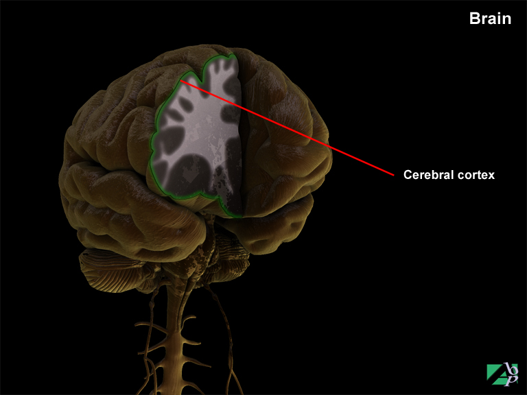
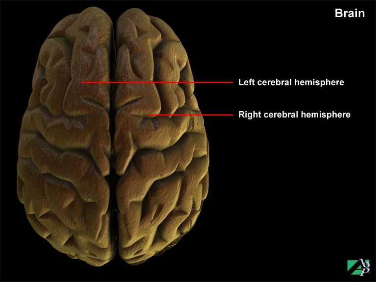
The cerebrum accounts for about 80 percent of total mass of the brain. It consists of the right and left cerebral hemispheres that are divided (though not completely) by a longitudinal fissure. Internally, portions of the two hemispheres are connected by a large tract of white matter called the corpus callosum. Each hemisphere fits into the dome of the cranial vault of the skull. The cerebrum comprises a surface layer referred to as the cerebral cortex that is composed of grey matter and below this, a thick layer of white matter. The cerebral cortex features numerous folds and grooves that are called convolutions which form during fetal development. These convolutions allow for a larger area of grey matter that is made up of nerve cell bodies. The elevated fold of the convolutions are called gyri and the depressed grooves are called sulci.
Cerebral Cortex
The cerebral cortex is about 1/2 inch in thickness, although this varies slightly in different areas of the brain. It is composed of what is referred to as grey matter and white matter.
The grey matter of the cerebral cortex is a densely packed region comprising neurons, dendrites and capillary blood vessels. The neurons (nerve cells) are the basic operative units of the brain. Below the grey matter is a thicker inner layer, the sub-cortical layer, which is white in appearance.
The white matter is made up of dendrites and myelinated axons. These neural fibers form the billions of connections within the brain by which information in the form of electrical impulses is transmitted to the selective areas of the brain. There are three types of fiber tracts within the white matter: association fibers, commissural fibers and projection fibers. Association fibers belong to a given hemisphere and only conduct neural impulses within their given hemisphere. Commissural fibers connect neurons from one hemisphere to another. Projection fibers form tracts that transmit and receive impulses from neurons in the cerebrum to other parts of the brain and spinal cord.
Cerebral Hemispheres
The cerebral hemispheres are divided by the sulci into large subdivisions called lobes. Four of the lobes are on the surface of the hemispheres and are named according to the overlying cranial (skull) bones. These lobes are the frontal lobe, parietal lobe, temporal lobe and occipital lobe. A fifth lobe, the insula, lies deep in the cerebrum and is covered by portions of the frontal, parietal and temporal lobes. The two hemispheres carry out different functions. Generally the left hemisphere carries out analytical and verbal functions. The right hemisphere is the source for spatial and artistic skills. The hemispheres, via the connection with the corpus callosum (white matter), share memory and learning.
Frontal Lobes
The frontal lobe is the anterior (front) portion of each cerebral hemisphere. The frontal lobes' functions include initiating voluntary motor impulses for the movement of skeletal muscles, analyzing sensory experiences and providing responses relating to personality. The frontal lobes also involve responses related to memory, emotions, reasoning, judgement, planning and verbal communication.
A highly specialized area of the frontal lobe is the motor speech area, known as Broca's area. Generally located in the left hemisphere, mental activity in Broca's area causes selective stimulation of motor impulses in motor centers elsewhere in the frontal lobe, which in turn cause coordinated skeletal muscle movement in the pharynx and larynx. At the same time, motor impulses are sent to the respiratory muscles to regulate air movement across the vocal cords. This combined muscular stimulation translates thought patterns into speech.
Parietal Lobes
The parietal lobe lies behind the frontal lobe. The cortex (grey matter) of the parietal lobes is concerned with the primary and secondary sensory functions and complex association mechanisms involved in higher-level integration and, in the dominant hemisphere, speech.
Somatosensory Function
Just behind the central sulcus or fissure of Rolando (a deep furrow that separates the parietal lobe from the frontal lobe) lies the somatosensory area for primary sensation in the postcentral gyrus of the parietal lobe. The body image is represented in the postcentral gyrus in an orderly sequence of dermatomal patterns (areas of skin supplied by branches of spinal nerves), paralleling that of the precentral motor cortex in the frontal lobe. Those body areas that have the most highly developed senses of touch and acuity have the largest representations in the sensory cortex (the thumb and forefinger areas, for example). This area receives fibers conveying sensation from the skin, muscles, joints and tendons of the opposite side of the body. Some of the sensations are primary; others are more complex.
The parietal lobe functions in the understanding of speech and in verbal articulation of thoughts and emotions. The parietal lobe also interprets the textures and shapes of objects as they are handled.
Temporal Lobes
The temporal lobe lies beneath the parietal lobe and the posterior portion of the frontal lobe separated from the frontal lobe lying above it by the lateral or Sylvian fissure and from the parietal lobe above and further back and the occipital lobe behind by smaller fissures.
The temporal lobe contains auditory centers that receive sensory fibers from the cochlea of the ear. It also interprets some sensory experiences and stores memories of both auditory and visual experiences.
During each part of the conscious waking state, the brain receives information. All information is stored and recorded in the temporal lobe of the brain to be recalled or remembered at a later date so that proper judgments can be made based on past experience.
The events preserved in memory however, mostly cannot be voluntarily recalled. Only memorable or emotional events or events that were deliberately rehearsed, learned and repeated afterwards, can be brought to conscious awareness.
Occipital Lobes
The occipital lobe forms the posterior portion of the cerebrum and has no distinct separation from the temporal and parietal lobes. Functionally, the occipital lobes are concerned with vision. The occipital lobe interprets eye movements by directing and focusing the eye. It is also responsible for visual association, correlating visual images with previous visual experiences and other sensory stimuli.
Insula
Little is known about the function of the insula though it primarily integrates other cerebral activities and may have some function with memory.
Limbic Lobes
These are older portions of the forebrain, which, during evolutionary expansion of the hemispheres, have become squeezed over to the middle, and deep aspects of the hemispheres.
The limbic system is a group of fiber tracts and nuclei that form a ring around the brain stem. They were once called rhinencephalon because of their importance in the central processing of olfactory (smell) information. These structures are now thought to be involved in basic emotional drives such as anger and fear.
Basal Ganglia
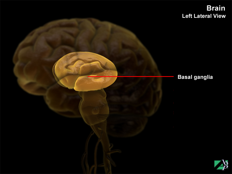
The basal ganglia are specialized paired masses of grey matter located deep within the white matter of the cerebrum. The most prominent of the basal ganglia is the corpus striatum. The corpus striatum is composed of several masses of nuclei. The caudate nucleus is the upper mass. A thick band of white matter lies between the caudate nucleus and the next lower two masses which is called the lentiform nucleus. This consists of a lateral portion named the putamen and a medial portion called the globus pallidus.
The basal ganglia control unconscious contractions of certain skeletal muscles such as those of the upper extremities involved in involuntary arm movements during walking. The globus pallidus regulates the muscle tone necessary for specific intentional body movements. Trauma or disease involving the basal ganglia generally causes a variety of motor movement dysfunction including rigidity, tremor and rapid and aimless movements.
Neurons (Nerve Cells)
A neuron or nerve cell contains a cell body with a nucleus. A number of fine branches, called dendrites, extend from the cell body. The dendrites carry impulses towards the cell body that are known as incoming impulses. The neuron also has a large single branch, the axon, which carries impulses away from the cell body. These impulses are known as outgoing impulses. The axon is covered by a white lipoid (resembling fat) called myelin, which acts as an insulator to prevent short-circuiting of neural impulses. The white matter substance of the brain is made up of bundles of these myelinated axons, called tracts.
Sensory (Afferent) Impulses
Incoming neural impulses are called afferent impulses or sensory nerve impulses. These sensory impulses are initiated at the peripheral receptor organs of the body: the skin, subcutaneous tissue, muscles, ligaments, internal organs and organs of the special senses. Afferent impulses occur in response to outside or environmental conditions and internal conditions within the body itself such as pain, heat, itching.
Motor (Efferent) Impulses
Outgoing impulses via the axons are known as motor impulses or efferent impulses. These nerve impulses are transmitted from the brain or spinal cord to a structure of the body such as a muscle or gland.
Electrical Transmission of Impulses
The membranes of neurons are polarized. This means there is an electrical potential difference between the inside and outside of the nerve cell due to unequal concentration of electrically charged chemical substances. Inside the unstimulated cell there is more potassium and less sodium. Stimulation of the neuron starts a wave of depolarization that creates a shift in polarity. Sodium moves into the cell membrane and potassium moves out. This shift is accompanied by electrical changes and the impulse spreads. Re-polarization occurs rapidly.
Brain Waves
Neurons within the cerebral cortex continuously generate electrical activity. These may be recorded by placing electrodes attached to precise locations on the scalp. This produces what is known as an electroencephalogram (EEG). The pattern of the EEG output is commonly called brain waves These brain waves are first emitted from the developing brain during early fetal development and continue throughout life. The cessation of brain wave patterns is indicative of death.
Certain brain wave patterns indicate healthy mental functions; abnormalities in these patterns are of clinical significance in diagnosing brain trauma and various diseases and also some mental illnesses. Normally there are four kinds of brain wave patterns; they are alpha waves, beta waves, theta waves and delta waves. Theta waves in an adult may be indicative of severe emotional stress while the presence of delta waves in a conscious individual is suggestive of brain damage.
Diencephalon
The diencephalon is a relatively small part of the brain situated between and below the cerebral hemispheres and above the medulla oblongata and the cerebellum. It is sometimes referred to as the between brain. It is divided into the epithalamus, subthalamus, hypothalamus and thalamus.
Thalamus
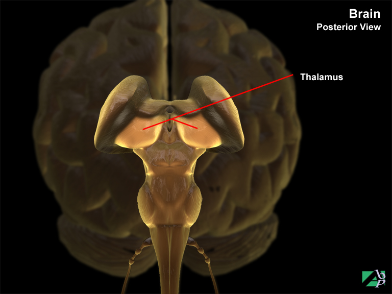
The thalamus constitutes nearly four-fifths of the diencephalon. It is a paired organ.
It is the chief relay center of sensory impulses travelling to the cortex of the brain. Sensory impulses from the body travel up the spine to the thalamus which then relays the sensation to the cerebrum for evaluation and response.
The thalamus also performs some sensory interpretation. The cerebral cortex discriminates pain and other tactile stimuli, but the thalamus responds to general sensory stimuli and provides crude awareness.
Hypothalamus
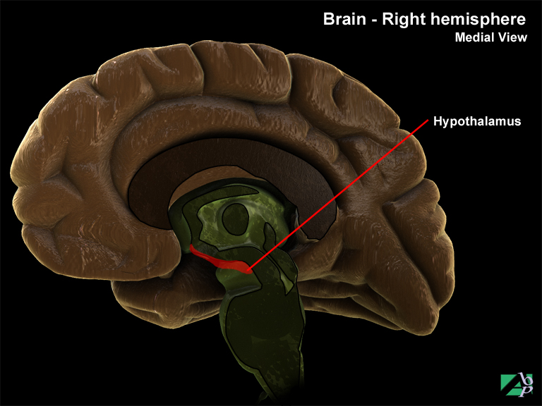
The hypothalamus is a small portion of the diencephalon located below the thalamus. It is connected with the pituitary gland that hangs below it in the sella turcica (cavity in the base of the skull) by means of a narrow stalk called the infundibulum.
The hypothalamus has many vital functions. It activates and controls the autonomic nervous system. It regulates body temperature, body fluid balance, sleep, perspiration, sexual activity and the development of secondary sex characteristics and emotional control. It has also been shown to be involved in the regulation of food intake although the exact mechanisms of this regulation are not yet clear.
Pituitary Gland
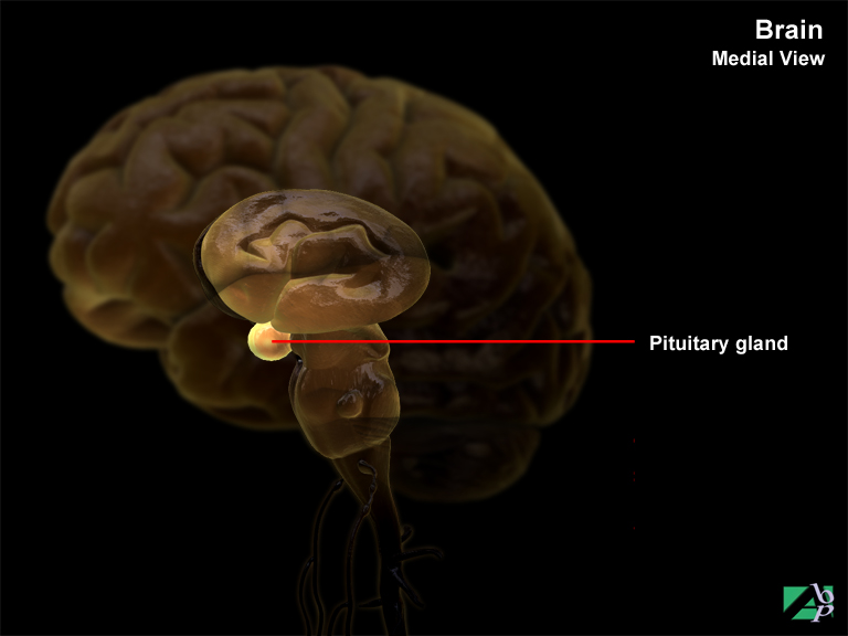
The pituitary gland is a small structure about the size of a pea. Its main function is the production of a number of hormones, among these being growth hormones that stimulate growth of the body and which also influence the metabolism of carbohydrates, fats and proteins. It also produces hormones that control the function of the thyroid and adrenal glands and hormones that stimulate the gonads, ovaries and testicles. In addition it stimulates the production of milk. Other hormones produced affect the contraction of the smaller blood vessels and the output of urine.
The Autonomic Nervous System
The autonomic nervous system refers to a system of peripheral nerves and ganglia whose neural activity is automatic and not subject to voluntary or conscious control. The central nervous system is concerned with the execution of voluntary or wilful acts and with the reception and interpretation of sensory impulses, however many of the functions of the body are independent of will. Glandular activity and the activity of organs such as the stomach or intestines are free from conscious act. These activities are managed by the autonomic nervous system.
The autonomic nervous system innervates the smooth muscles of the body such as those found in the stomach, gallbladder, arteries, the glands and the heart. It controls without intervention of the central nervous system most of the internal structures of the body: the eyes, saliva glands, arteries, windpipe, lungs, heart and liver to name just some.
The autonomic nervous system is divided into two parts, the sympathetic nervous system and the parasympathetic system. The difference in these two systems is in the manner in which they affect the organs or tissues they control. Generally the sympathetic nervous system acts as a stimulator and the parasympathetic system as an inhibitor or depressor.
Another difference in the two systems is the part of the central nervous system with which each of the systems communicates. The parasympathetic system communicates with the midbrain, medulla and lower part of the spinal cord. The sympathetic system communicates with the spinal cord only.
Epithalamus
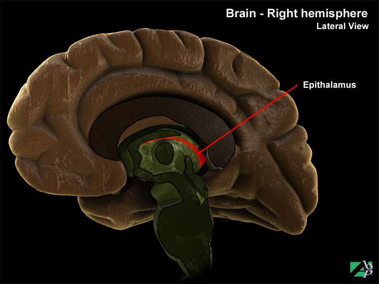
The epithalamus is the dorsal portion of the diencephalon. The roof of the epithalamus consists of a vascular choroid plexus where cerebrospinal fluid is produced.
Brain Stem
The brain stem comprises the midbrain called the mesencephalon and the hindbrain, the rhombencephalon. The latter is made up of the pons, medulla oblongata, reticular activating system and the cerebellum. With the exception of the cerebellum, it is tubular in shape and extends from the diencephalon above and in front to the lower end of the medulla below and behind. It forms a conduit for fiber tracts from the spinal cord below to the higher brain centers above.
Pons
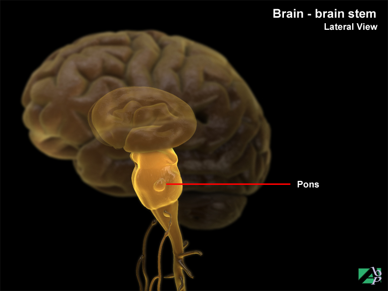
The name pons is derived from the Latin word "pons" meaning "bridge". It is the crossover point for ascending sensory and descending motor tracts linking higher and lower centers, and for transverse fiber pathways entering and leaving the cerebellum on each side.
Its significance lies in the fact three cranial nerves arise from it and along with the medulla oblongata it regulates the rate and depth of breathing. The cranial nerves are the sixth cranial nerve, the abducens nerve, the seventh cranial nerve, the facial nerve the eighth cranial nerve and the vestibuloocular nerve.
Medulla oblongata
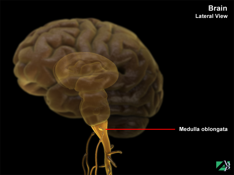
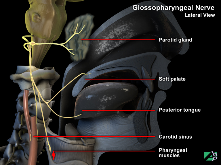
The medulla oblongata or, as it is more often called, the medulla, is a bulbous structure that resembles the spinal cord and is continuous with the pons above and the spinal cord below. The medulla comprises vital nuclei and white matter that form the descending and ascending communication tracts between the spinal cord and the brain. Various cranial nerves arise from the medulla including the glossopharyngeal nerve, accessory nerve, hypoglossal nerve and vagus nerve. Other nuclei within the medulla function as autonomic centers for controlling vital visceral functions including heartbeat, arterial blood pressure and rate and depth of breathing.
Reticular Activating System (RAS)
This is a complex network of nuclei and nerve fibers that function to arouse the cerebrum. Portions of this network are located in the spinal cord, pons and midbrain, thalamus and hypothalamus. The principle functions of the reticular activating system are to keep the cerebrum in a state of alert consciousness and to selectively monitor sensory impulses perceived by the cerebrum. The reticular activating system also aids the cerebrum to maintain contraction of skeletal muscles.
Meninges of the Brain
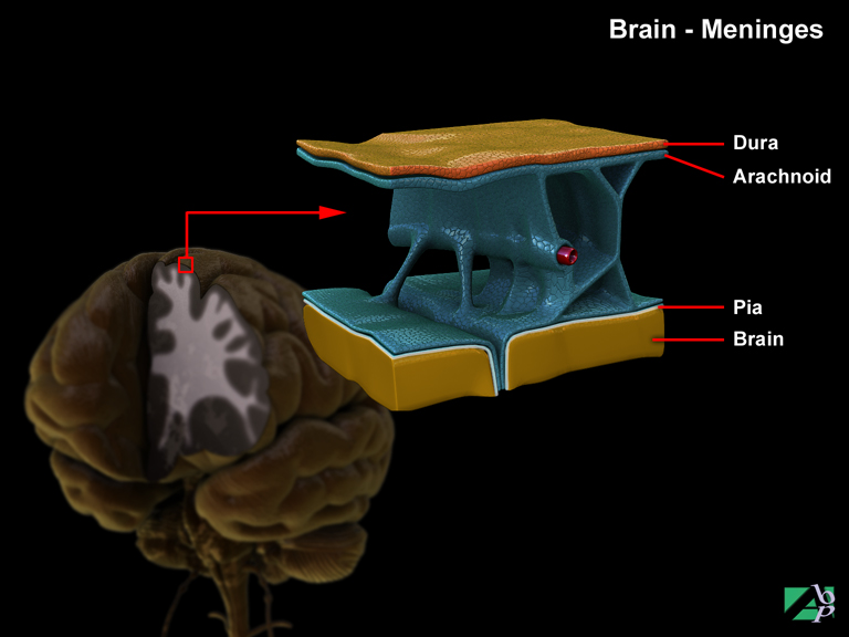
Three membranes called meninges enclose the brain and spinal cord. These form a continuous protective three-layered sac separating brain and spinal cord from overlying bone. These membranes are the pia mater (inner membrane), the arachnoid membrane (middle membrane) and the dura mater (outer membrane).
Pia Mater
The pia mater or pia, is a thin vascular membrane that is attached to the surface of the brain and spinal cord. It is highly vascularized and functions to support the small blood vessels that nourish the underlying cells of the brain and spinal cord.
Arachnoid Membrane
The arachnoid or middle membrane is a thin transparent membrane that lies between the pia and the dura. The arachnoid is separated from the pia mater by a shallow space called the subarachnoid space. This space contains cerebrospinal fluid (CSF).
Dura Mater
The dura mater (or simply dura) is in contact with bone and is composed primarily of tough fibrous connective tissue. The dura is made up of two layers: the endosteal or periosteal layer that adheres lightly to the inner surface of the skull bones, and the meningeal layer that follows the general contour of the brain. These two layers stick to each other over most of the brain except in certain locations where the layers separate to enclose the cranial dural sinuses. In the spine there is no connection between the dural sheath and the vertebra, rather there is a potential cavity called the epidural space.
In certain locations within the skull, the inner layer of the dura becomes reduplicated. These reduplications project into the cranial cavity and form partitions. The major partitions formed by these reduplications or infolding of the inner layer of dura are: the falx cerebri, the falx cerebelli, the tentorium cerebelli and the diaphragma sellae.
The Falx Cerebri
The falx cerebri is formed by an infolding or reduplication of the inner layer of dura in the midline, above, and extends downward to form a partition separating the two cerebral hemispheres.
Diaphragma Sellae
The diaphragma sellae is a small dural infolding which covers the sella turcica (depression in the base of the skull). In its posterior portion is a small opening through which the infundibulum or stalk from the hypothalamic region of the brain above reaches the posterior lobe of the pituitary gland which lies in the hollow of the sella below.
Tentorium Cerebelli
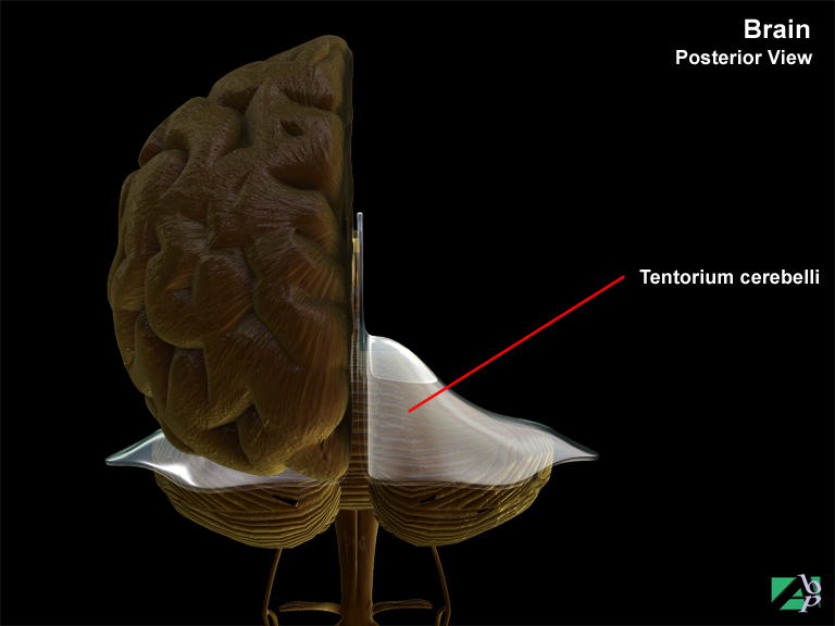
The tentorium cerebelli (often called simply the tentorium) is an important structure because of its location and anatomical relationships. The tentorium supports the occipital lobes of the cerebrum. It is attached at the sides and behind to the inner surface of the occipital bone, and in front to the petrous temporal bone. The anterior border of the tentorium is a free edge, concave in shape, and surrounding a large midline opening, the incisura tentorii or notch. The brain stem passes through this opening that is important in terms of trauma. Sudden violent movement of the brain from an impact to the head may throw the brain against the sharp free edge of the incisura, causing brain contusion or laceration. Additionally, when there is swelling or edema of the brain with increased intracranial pressure, the under side of the medial portions of the hemispheres may herniate (transtentorial herniation) through the opening of the tentorium, compressing the brain stem.
Ventricular System
Cerebral ventricles are cavities or chambers within the brain. These are the lateral ventricles (there are two), the third ventricle and the fourth ventricle.
The ventricles of the brain are where the cerebrospinal fluid is produced. The fluid is formed by tissue known as the choroid plexus that exists in each ventricle.
The lateral ventricles are found in the right and left cerebral hemispheres of the cerebrum. Each lateral ventricle consists of a central part and three horns.
The third ventricle is situated between the thalamus and the hypothalamus. This ventricle communicates with the lateral ventricles through an opening called the interventricular foramen. There are two such openings, one to communicate with the right lateral ventricle and the other to communicate with the left lateral ventricle.
The fourth ventricle is situated between the pons in front and the cerebellum in the back. It communicates with the third ventricle through a channel called the aqueduct of Sylvius. It also communicates below with the central canal of the spinal cord.
Cerebrospinal Fluid (CSF)
(Show Animation)
Cerebrospinal fluid is a clear colorless fluid consisting of glucose, urea, proteins and salts in addition to some white blood cells.
Cerebrospinal fluid circulates continuously around the brain where it acts as a cushion against jarring or sudden shocks. It also circulates nutritive substances filtered from the blood. The formation of cerebrospinal fluid is continuous and the direction in which it circulates is from top to bottom i.e. from the lateral ventricles down through the third and fourth ventricles and into basal cisterns. The basal cisterns are dilated portions of the subarachnoid space at the base of the brain.
The cerebrospinal fluid is eventually absorbed back into the general venous circulation by way of the arachnoid villi that project into the various venous sinuses of the brain. This formation and absorption rate results in a fairly constant volume of circulating fluid.
If there is increased production, decreased absorption or blockage of flow, an increased accumulation of fluid within the ventricles will result and create intracranial pressure.
Cerebellum
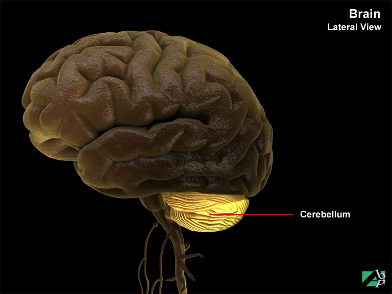
The cerebellum (sometimes referred to as the little brain) is the second largest structure of the brain and occupies the inferior and posterior aspects of the cranial (skull) cavity.
The cerebellum consists of two hemispheres and a central constricted area called the vermis. Like the cerebrum, the cerebellum has a thin outer layer of grey matter called the cerebellar cortex and a thick deeper layer of white matter. Three paired bundles of nerve fibers called cerebellar peduncles support the cerebellum and provide it with tracts for communicating with the rest of the brain.
The principle function of the cerebellum is coordinating skeletal muscle contractions. Impulses for voluntary muscle movement originate in the cerebral cortex and are coordinated by the cerebellum The cerebellum constantly initiates impulses to selective motor units for maintaining posture and muscle tone.