Medical Glossary - Letter L
This medical glossary of terms beginning with the letter "L" contains the more common medical terms one might expect to encounter in a medical report or in hospital notes. The glossary is intended as a quick reference only; many of the terms are also referenced and illustrated in more detail in the medical libraries, to which you should refer for more detailed information.
Labial Fold
The groove in the mouth between the gums and the lip
Lacrimal Bones
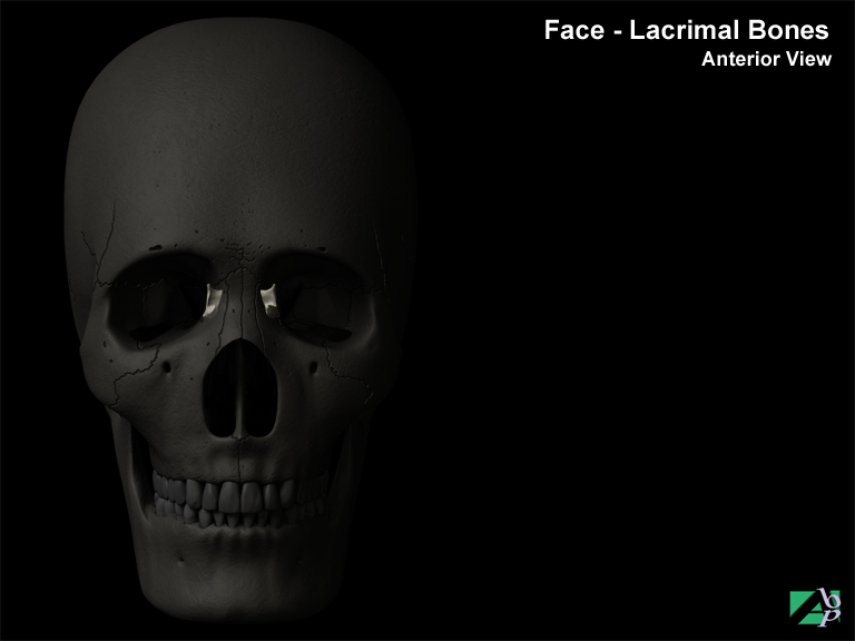
These are small thin bones that form the anterior part of the medial wall of each orbit. The medial wall is the wall closest to the nose. It is joined with the frontal and ethmoid bones of the skull and with the maxilla (upper jaw bone). They each have a groove that acts as a canal for tears from the eye to drain into the nasal cavity
Lacrimal Canaliculi
The lacrimal canaliculi also known as the lacrimal ducts are the two tear carrying ducts that carry the tears from the eye to the nasolacrimal duct, which carries the tears into the nose. They are located near the inner angle of the eyelids (towards the nose)
Lacrimal Punctum
The lacrimal punctum or puncta lacrimalia are tiny openings on the inner surface of the eyelids (one on each eyelid) near the nose through which the tears produced by the lacrimal gland are drained. The tears drain through the punctum into the lacrimal canaliculus (a canal through which the tears enter the nose)
Lacrimal System
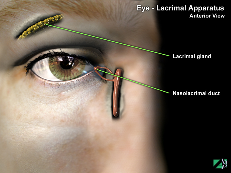
This system consists of a gland (lacrimal gland) and a series of ducts (lacrimal ducts) that drain secretion of tears. The lacrimal gland is located in the upper portion of the orbit. It secretes lacrimal fluid (tears) into the conjunctival sac of the upper eyelid. This fluid serves to lubricate the eyelid and eye and also reduces the chance of infection. In response to irritating substances coming into contact with the conjunctiva the gland becomes stimulated to over secrete, which serves to wash the eye
Lamboidal Suture
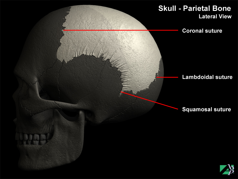
The skull is divided into regions by tissue and cartilage called sutures; the lamboidal suture divides the parietal and temporal bones
Lamellar Keratoplasty
Lamellar keratoplasty involves removal of the lamellae (the outer corneal layer) and replacement with normal corneal tissue from the patient (autograft)
Laminae
Part of the neural arch of a vertebra
Laminectomy
A spinal operation in which the posterior arch of a vertebra (the lamina) is surgically removed
Laminotomy
Laminotomy is a partial laminectomy. In this procedure only part of the lamina is removed. The advantage of this procedure over laminectomy is it does not compromise the structural integrity of the vertebra
Laparoscopy
Laparoscopy is the visualization of the interior of the abdomen using a laparoscope. A laparoscope is a tubular surgical instrument with optical lenses and a lighting system. It has advantages over open surgery in that only a small incision is required in the abdominal wall; it may be for investigation purposes or to perform surgery on some intra-abdominal structure
Laparotomy
A laparotomy is the surgical opening of the abdomen. It is often the surgical approach for some other procedures. It is also often performed as an exploratory procedure where it is suspected some internal organ has suffered serious damage or internal bleeding is suspected and vital signs are affected
Large Intestine
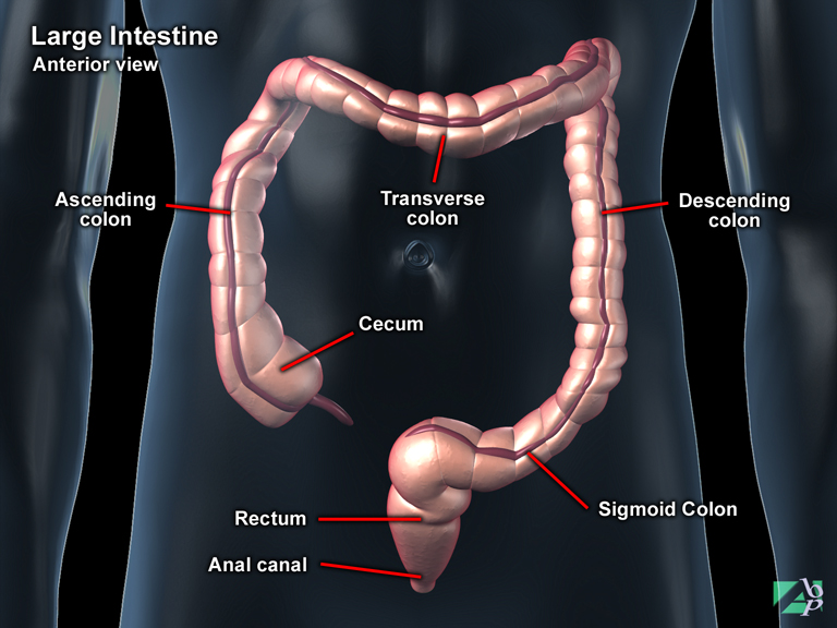
The large intestine is so named not because it is longer than the small intestine (it isn't) but because it is larger in diameter. The large intestine is attached to the abdominal wall by a specialized portion of the abdominal mesentery known as the mesocolon. Like the small intestine, the large intestine has a number of divisions, they are: the caecum, colon, rectum and anal canal. The large intestine begins at the end of the ileum (the terminal end of the small intestine) and rises to just below the liver where it crosses over to the left side of the lower abdomen and descends into the pelvis where it terminates at the anus. The large intestine plays no role in the digestion of food although it does absorb water and electrolytes (irons). It serves to form, store and expel waste matter from the body. The largest section of the large intestine is the colon, which consists of four segments, the ascending colon, transverse colon, descending colon and sigmoid. The first three segments simply describe the direction the colon traverses and the latter (sigmoid) describes the "S" shape taken by the terminal end of the colon. The first portion, the caecum is a dilated pouch that hangs behind a membrane known as the ileocecal valve, which serves to prevent the backflow of digested material. At the terminal end of the sigmoid is the rectum and anal canal; the anus is the external opening of the anal canal
Laryngeal
Referring or pertaining to the larynx
Laryngopharynx
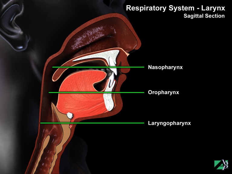
The laryngopharynx is a portion of the pharynx that extends from the hyoid bone to the esophagus at which point the respiratory and digestive systems become distinct. Food is passed to the esophagus and air to the trachea
Laryngoscopy
Laryngoscopy is the visual inspection of the interior of the larynx (voice box). A special instrument called a laryngoscope comprising lenses and lights is used to perform the examination
Larynx
(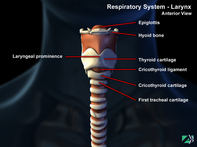
The larynx or voice box joins the pharynx to the trachea. It is approximately in the middle part of the neck, in line with the 4th, 5th and 6th cervical vertebra. The larynx serves two purposes. One is to prevent food and fluid from entering the lungs during swallowing and to allow air through when breathing. The other is to produce sound. The larynx is made up of a framework of cartilages, nine in all. The principal cartilages are the thyroid cartilage (the Adams apple), the epiglottis and the cricoid cartilage. The remaining cartilages form attachment points for the vocal cords. The vocal cords are formed by two bands of connective tissue that stretch from the front to the rear of the larynx. They vibrate when air flows out from the lungs. Changes in the tension of the vocal cords result in a variation in the pitch and quality of the voice. The laryngeal intrinsic muscles regulate tension of the vocal cords. Another group of muscles, the extrinsic muscles, are responsible for elevating the larynx to prevent food from entering during the act of swallowing
Lateral
Refers to the outer side, away from the midline point of the body
Lateral Collateral Ligament
(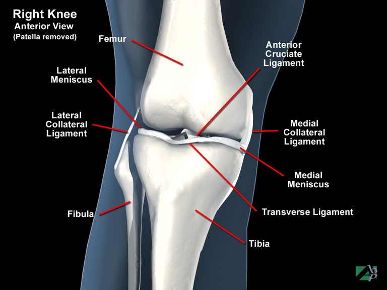
A knee ligament
Lateral Femoral Cutaneous Nerve
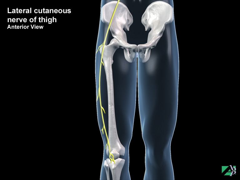
This is a sensory nerve only. It supplies sensation to the skin of the back and outside of the thigh and the rear of the thigh
Lateral Plantar Artery
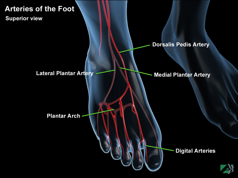
The lateral plantar artery commences in the ankle and supplies the sole of the foot, in the foot it joins with the dorsal pedis artery from which the digital arteries arise
Lateral Rectus
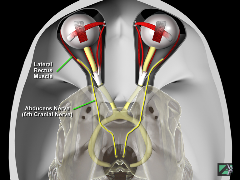
An eye muscle originating within the orbit and inserting onto the sclera. It abducts the eye. The abducent nerve, a cranial nerve, innervates it
Lateral Ventricles
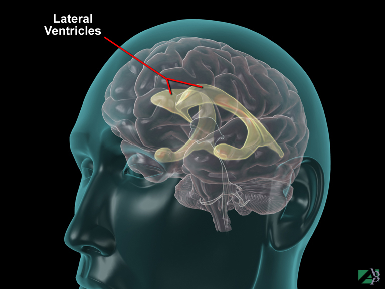
The lateral ventricles are chambers in the left and right hemispheres of the brain where cerebrospinal fluid is produced
Latissimus Dorsi
A muscle in the back that originates from the spinous process of the lower thoracic, lumbar and sacral vertebra and extends around the trunk and inserts onto the bicipital groove of the humerus. It assists extension, adduction and medial rotation of the arm
LeFort Fracture
Fracture of the maxilla. Maxilla fractures are classified as LeFort fractures of which there are three types: LeFort 1 is the most frequent type. It is a transverse fracture occurring just above the level of the teeth in the alveolar process. This fracture results in the roof of the mouth (the hard palate) separating. LeFort 11 is referred to as a pyramid fracture, as the fracture lines resemble the shape of a pyramid. In this vertical fracture, the fracture block contains the frontal processes of the maxilla and the nasal bones and is often associated with a depressed or comminuted fracture of a zygoma. The fractures also commonly extend through the lacrimal bones and the floor of the orbit. LeFort 111 is the most complex of all facial fractures as it involves the entire middle third of the face. The fractures extend from the nasofrontal junction down the medial wall of the orbit, across the floor of the orbit and through the maxilla, ethmoid and sphenoid bones, with complete separation of the middle facial skeleton
Left Hypochondriac Region of Abdomen
The left hypochondriac region contains the spleen, portions of the right kidney and the small intestine. It describes the location of the left upper one third of the abdomen
Left Inguinal Region of Abdomen
The left inguinal region contains some of the small intestine, the descending colon and the sigmoid colon. It describes the location of the left lower one third of the abdomen
Left Lateral Region of Abdomen
The left lateral region contains the descending colon, some of the left kidney, and the small intestine. It describes the location of the left lateral one third of the abdomen
Lens
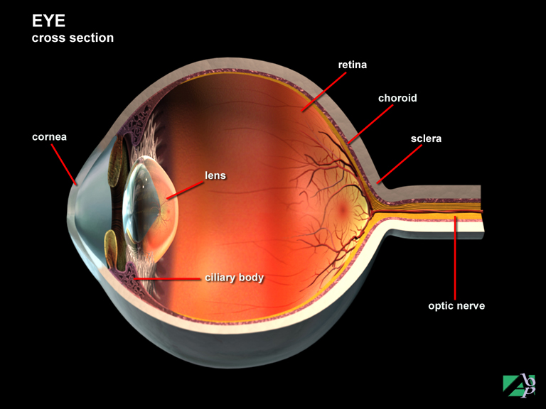
The lens lies within the eyes interior between the smaller aqueous chamber and the larger vitreous chamber. Its purpose is to focus light images on the retina for transmission to the brain for dissemination
Lentiform Bone
Another name for the pisiform bone, one of the carpal bones of the wrist
Levator Labii Superioris
A facial muscle originating from the maxilla and zygoma, it serves to raise the upper lip
Levator Palpebrae Superiores
A facial muscle that originates from the sphenoid bone of the skull and inserts onto the skin of the upper eyelid. It elevates and retracts the eyelid. It is innervated by the oculomotor nerve, a cranial nerve
Levator Scapulae
A muscle in the upper back originating from the vertebral transverse processes of C1-C4, which inserts onto the border of the scapula. It raises the scapula
Levatores Costarum
A muscle in the middle of the back originating from the transverse processes of C7-T11, which inserts onto the posterior surfaces of the ribs. It elevates the ribs
Ligamentum Patellae
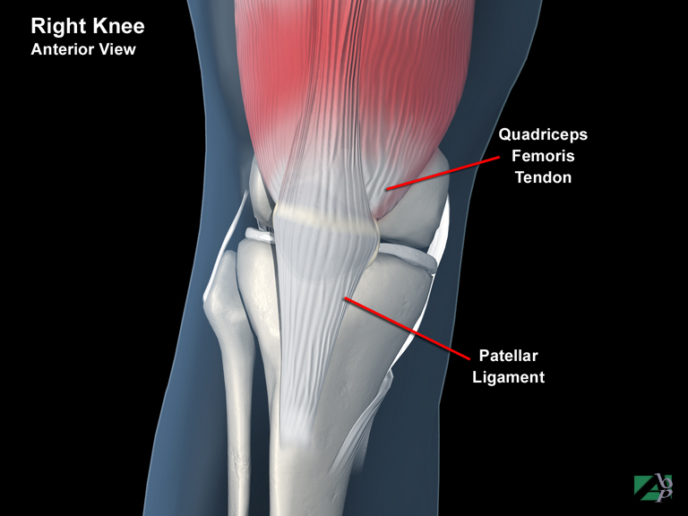
A ligament that supports the knee
Ligamentum Teres
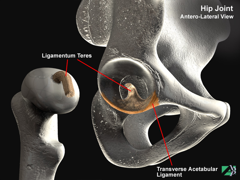
A ligament that supports the hip joint
Ligation
To tie off, as with an artery or vein to stop the bleeding
Limbic Lobe
These are older portions of the forebrain, which during evolutionary expansion of the hemispheres have become squeezed over to the middle and deep aspects of the hemispheres
The limbic system is a group of fiber tracts and nuclei that form a ring around the brain stem. They were once called rhinencephalon because of their importance in the central processing of olfactory (smell) information. These structures are now thought to be involved in basic emotional drives such as anger, and fear
Linear Skull Fracture
A straight single-line fracture with no bony fragments or displacement. Most linear skull fractures occur in the parietal region of the skull and extend down to the base of the skull
Lingual
Pertaining or referring to the tongue
Lingual Frenectomy
Lingual frenectomy is the surgical excision of the frenulum, which is the membrane that connects the underside of the tongue with the floor of the mouth
Lingual Frenotomy
Lingual frenotomy is the surgical division of the frenulum, which is the membrane that connects the underside of the tongue with the floor of the mouth
Liver
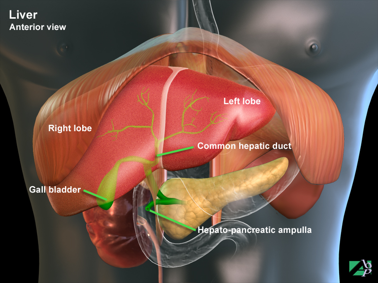
The liver is the largest of the body's internal organs and in an adult weighs about 1.3 kilograms. It is located in the upper region of the abdomen below the diaphragm and spans the entire width of the abdominal cavity from front to rear. The lower regions of the liver are in contact with the right kidney and the stomach and colon. It is attached to the abdominal wall and held by ligaments. The liver's general purpose is filtration and storage of blood, secretion of bile, excretion of bile pigment and general detoxification. The liver consists of four lobes of unequal size and shape. The basic cells of the liver are called hepatic cells. One of the primary functions of these cells is the production of bile, which is stored in the gallbladder and released into the small intestine. Bile salts emulsify fats, which then allow the fats to be digested by the intestinal enzymes produced by the pancreas. The hepatic cells also assimilate carbohydrates and proteins. They convert glucose to its stored form of glycogen and assist in maintaining a proper level of glucose in the blood. The liver also clears the blood of harmful compounds by removing them from the blood and secreting them into the intestine with the bile. The liver also processes the end products of fat digestion and fatty acids into cholesterol and other substances used by the body. Excess carbohydrates and protein are converted into fat by the liver. Digested proteins in the form of amino acids are broken down further by the liver into glycogen and other compounds. Some essential components of blood are manufactured by the liver, including most of the plasma proteins and blood clotting substances. Additionally the liver stores important vitamins and minerals including vitamins A, D, K and some group B vitamins
Longus Capitus
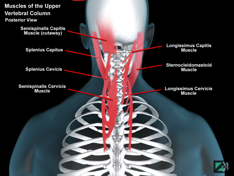
A muscle in the back of the neck that originates from the transverse processes of C3-C6 and inserts onto the occipital bone of the skull. It assists flexion of the cervical spine and the atlantooccipital joint
Longus Colli
A muscle in the back of the neck that originates from the transverse processes of C3-C7 and inserts onto the arch of C1 and the bodies of C2-C4. It assists flexion and rotation of the cervical spine
Lordosis
An abnormal forward curvature of the spine
Low Amplitude Force
A type of osteopathic manipulation
Lower GI Series
An x-ray of the colon
Lower Jaw
The mandible
Lumbago
A term occasionally used to describe low back pain
Lumbar
Pertaining or referring to the lumbar spine region
Lumbar Plexus
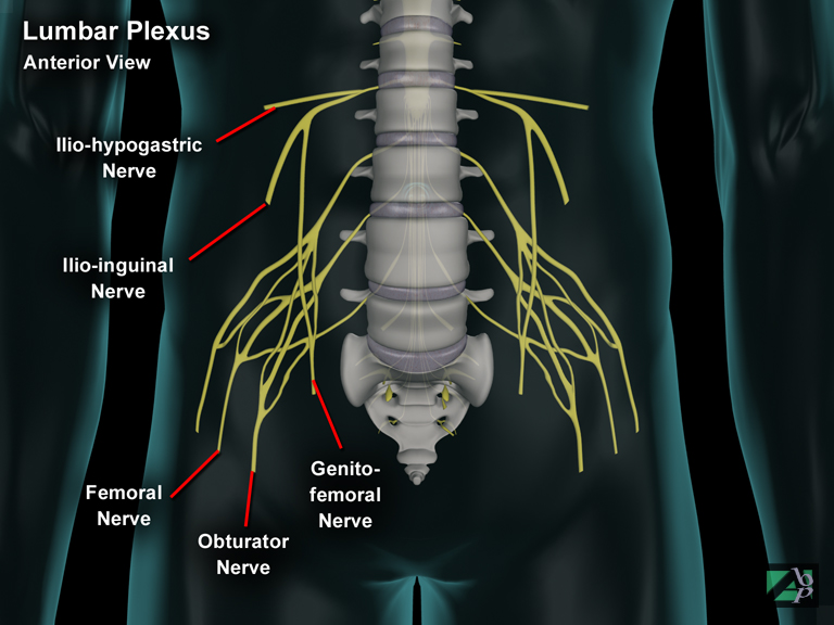
The lumbar plexus is formed by the spinal nerves arising from L1 though L4.
Peripheral nerves arising from this plexus innervate structures of the lower abdomen and anterior and medial portions of the lower extremity. These include the iliohypogastric nerve, ilioinguinal nerve, genitofemoral nerve and the obturator nerve
Lumbosacral Joint
The lumbosacral joint is the articulation between the 5th lumbar vertebra and the sacrum
Lumen
The hollow space within an artery or organ
Lunate
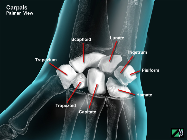
One of the carpal bones of the wrist, a moon shaped bone adjacent to the scaphoid. It has connections with the radius, and adjacent scaphoid and triquetrum and with the capitate and hamate in front
Lungs
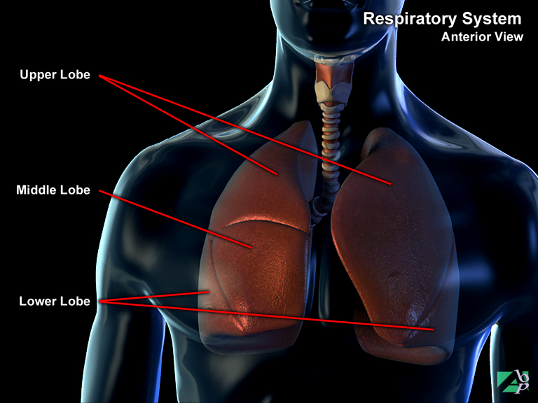
The paired lungs lie between the rib cage and diaphragm and are separated from each other by the heart and mediastinum (a space which contains the heart, esophagus, trachea and vital blood vessels e.g. aorta, pulmonary artery). The left lung is smaller than the right comprising a superior and inferior lobe. The right lung is comprised of three lobes, superior, middle and inferior. The lobes contain the alveoli. Membranes called pleura surround the lungs, one membrane is called the visceral pleura and another is the parietal pleura. The visceral pleura lines the outer surface of the lungs. The parietal pleura lines the thoracic walls and the diaphragm. The space created by the two pleura is called the pleural cavity. The pleura serve a number of important functions. They act as a lubricant to allow the lungs to move along the chest wall, they provide compartmentalization of the thoracic organs and also provide pressurization for the lungs
Lymphangiogram
A lymphangiogram is a radiological diagnostic test used to evaluate the functionality of the lymphatic system. The lymphatic system is a one-way circulation that channels tissue fluid, called lymph back to the heart. The lymphatic system may become clogged through disease, infection or through injury. The procedure is performed by injecting a blue dye into the hand or foot. The lymphatic system picks up the dye and circulates the dye throughout the lymph system. When the lymph system becomes visible an opaque material is then inserted into the system and x-rays are taken as it circulates through the system. This procedure can take hours to perform