Medical Glossary - Letter I
This medical glossary of terms beginning with the letter "I" contains the more common medical terms one might expect to encounter in a medical report or in hospital notes. The glossary is intended as a quick reference only; many of the terms are also referenced and illustrated in more detail in the medical libraries, to which you should refer for more detailed information.
Iatrogenic
This term describes a state, condition or occurrence caused by the treatment or advice given by a physician
Idiopathic
Not of traumatic origin, the term describes a condition or state that occurs spontaneously, or without any known cause
Ileitis
Inflammation of the ileum, which is part of the small intestine
Ileocecal
Refers to the area where the ileum and the caecum connect. The ileum is the last section of the small intestine; the caecum is the first section or beginning of the large intestine
Ileostomy
An operation whereby an incision or opening is made into the ileum along with an opening in the abdominal wall through which the contents of the large bowel may be discharged
Ileostomy Bag
A sac or bag attached to the abdomen to capture the discharge of fecess
Ileum
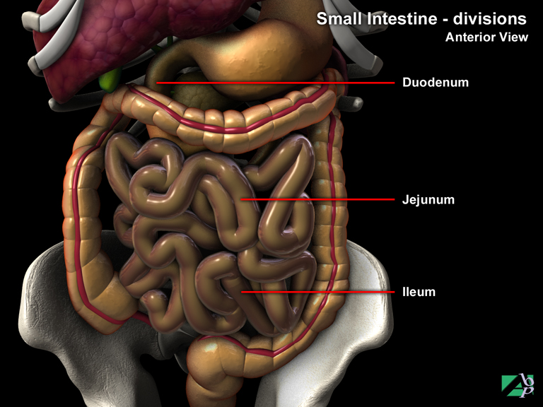
The last part of the small intestine
Iliac
Relating to the hip, the ilium, the top of the hip (pelvic) bone
Iliac Artery
This is a branch of the common iliac artery and supplies blood to the pelvis, buttock and the inner part of the thigh
Iliac Crest
The upper border of the ilium (part of the pelvis), it is from this area that bone is most often taken for bone grafts
Iliacus
A muscle originating in the abdomen and inserting onto the lesser trochanter of the femur. It assists flexion and medial rotation of the hip. The femoral nerve innervates it
Ilioinguinal Nerve
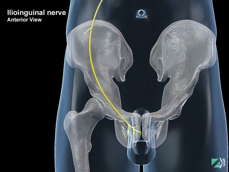
This is a sensory nerve that supplies sensation to the back of the thigh and genital area. It is rarely injured. It also innervates some abdominal muscles
Ilium
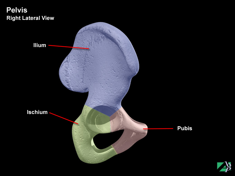
The upper portion of the pelvis
Impacted Fracture
A fracture where one end of the fractured bone is driven into the other end
Impermeable
Resistant
Incisional Hernia
Incisional hernias result from prior surgery where the surgery has left scarring or a weakness in the abdominal wall and the herniation occurs at or near the surgical site. Incisional hernias are common and sometimes the result of poor postoperative closure techniques when too much tension is placed on the surgical sight. In other cases obesity and other pre-existing conditions may pre-dispose the individual to hernia
Incontinence
Inability to control the discharge of urine
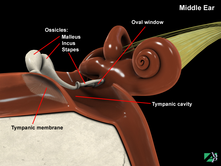
Incus
One of the three tiny bones (ossicles) of the middle ear
Indwelling Catheter
A catheter passed through the urethra into the bladder to drain urine, which is left in-situ for some period of time
Inferior Gluteal Nerve
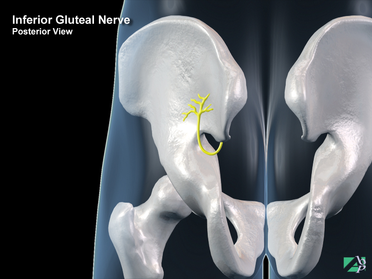
This nerve is found in the buttock. It provides motor activation to the extensor muscles of the thigh
Inferior Mesenteric Artery
The inferior mesenteric artery is an abdominal artery and is a branch of the abdominal aorta. It supplies blood to the transverse colon, descending colon, sigmoid and the rectum
Inferior Oblique
An eye muscle originating from the orbital margin and inserting onto the sclera. It elevates the eye. It is innervated by the oculomotor nerve, one of the cranial nerves
Inferior Rectus
An eye muscle originating from within the orbit and inserting onto the sclera. It depresses the eye. It is innervated by the oculomotor nerve, a cranial nerve
Inferior Vena Cava
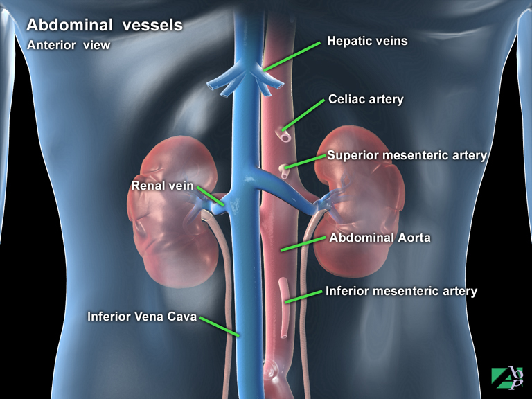
The inferior vena cava is a large vein, which receives blood from the lower extremity, abdominal organs and urinary and reproductive organs to empty into the right atrium of the heart (one of the chambers of the heart). It begins at the level of the fifth lumbar vertebra and travels through the diaphragm into the right atrium
Infraorbital Nerve
The infraorbital nerve, a sensory nerve is a branch of the maxillary nerve; its distribution is to the cheek, nostril, upper lip, upper teeth and lower eyelid
Infraorbital Region
The part of the face adjacent to the nose and below the eye socket
Infrapatellar Bursitis
Inflammation of the infrapatellar bursa, which is a bursa of the knee situated between the upper end of the tibia and the patella ligament
Infrascapular
Refers to the back region below the shoulder blade
Infraspinatus Muscle
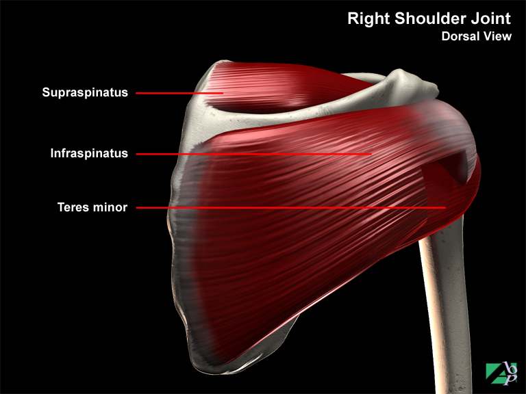
A muscle in the back of the shoulder, it aids flexion and rotation of the shoulder
Inguinal
Refers to the groin area
Inguinal Hernia
A hernia in the region of the groin, it is the most common occurring type of hernia
Inner Ear
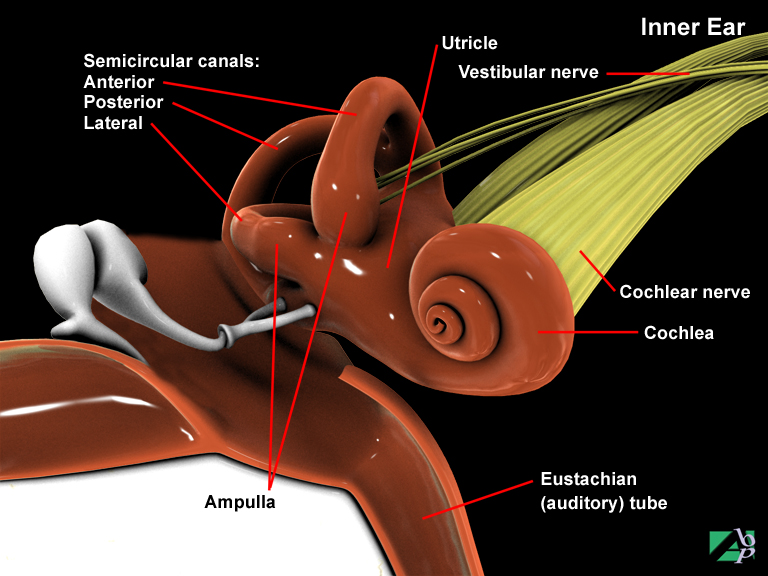
The innermost compartment of the ear is composed of two systems, the membranous and bony labyrinths. The bony labyrinth is formed from part of the temporal bone. It is structurally and functionally divided into three main regions: the cochlea, a snail-shaped tube which houses the organs of Corti (the end organ of hearing that is attached to the acoustic nerve); the semicircular canals, which contain part of the mechanism of balance; and the vestibule, the central region of the bony labyrinth that forms the connection between the semicircular canals and cochlea. The membranous labyrinth is a system of canals that lie within and congruent to the bony labyrinth and are filled with a viscous fluid known as endolymph. The space separating the membranous labyrinth from the walls of the surrounding bony labyrinth is filled with another, distinct fluid called perilymph. The inner ear is also responsible for equilibrium
Innominate Bone
Un-named, the name given to the hip bone, which forms part of the pelvis
Inorganic
Matter that is neither animal nor vegetable
Inspiration
To draw in breath
Instep
The arched upper surface of the foot from the ball of the foot (just behind the toes) to the heel
Integumentary
Pertaining to the skin
Interclavicular Ligament
A ligament that supports the sternoclavicular joint
Intercondylar
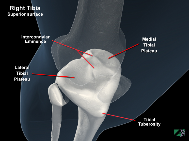
Between two condyles, which are bony protuberances at the lower end of some bones
Intercostal
Between the ribs
Intercostal Artery
There are two intercostal arteries, the arteria intercostalis suprema and the arteria intercostalis posteriores. The intercostalis suprema supplies blood to the 1st and 2nd intercostal spaces, vertebral column and some back muscles. The intercostalis posteriores supplies blood to the remaining intercostal spaces, the spinal column and cord and the muscles and skin of the back
Intercostal Catheter
An intercostal catheter is a drainage tube placed in the intercostal region, which is the space between adjacent ribs. It may be referred to simply as a chest tube
Intermediate Coup Lesion
A type of brain contusion where the contusion to the brain occurs near or around the impact to the skull
Internal Carotid Artery
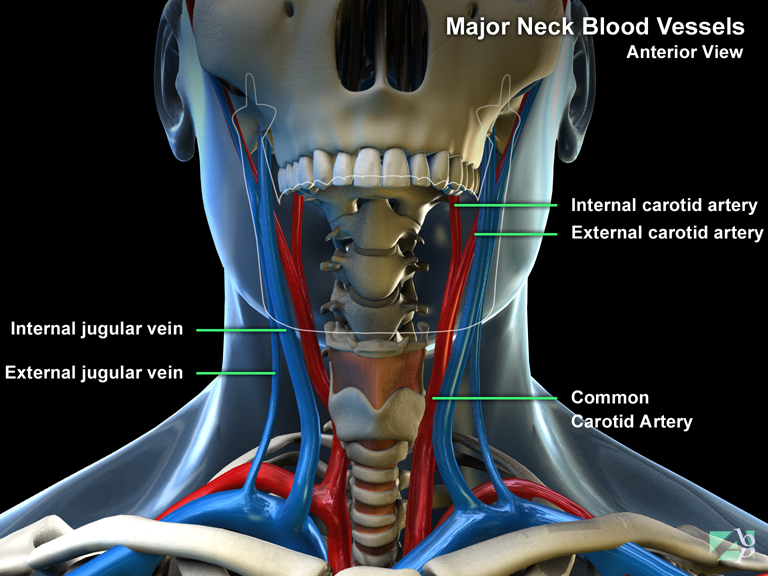
The internal carotid artery, like the external carotid artery is a branch of the common carotid artery. The internal carotid artery supplies blood to the head
Internal Intercostals
A muscle of the chest, originating from the inferior border of the ribs and inserting onto the superior border of the ribs. It aids respiration by elevating the ribs. The intercostal nerve innervates it
Internal Jugular Vein
The internal jugular vein is one of two veins on each side of the neck; the other is the external jugular vein. The internal jugular is the larger of the two and it drains blood from the brain, parts of the face and neck into the subclavian artery with which it unites to empty blood into the superior vena cava
Interosseous
Between two bones
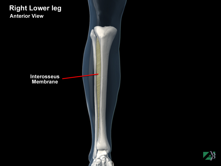
Interosseus Membrane
A membrane that extends between two bones, it is found in the leg and arm. In the arm it binds the radius to the ulna and in the leg the tibia to the fibula
Interossei Dorsal of Hand
A muscle on the back of the hand originating from the shafts of each metacarpal and inserting onto the proximal phalanges of the index and middle fingers. It assists flexion and abduction of the fingers. The ulnar nerve innervates it
Interossei Dorsal of Foot
A muscle on the top of the foot originating from the shafts of all metatarsals and inserting onto the 2nd, 3rd and 4th toes. It assists abduction of the toes. The lateral plantar nerve innervates it
Interossei Palmar
A muscle in the palm of the hand originating from the shafts of the 1st, 2nd, 4th and 5th metacarpals and inserting onto the thumb, index, ring and little fingers. The ulnar nerve innervates it
Interossei Plantar
A muscle in the sole of the foot originating from the shafts of the 3rd, 4th and 5th metatarsals and inserting onto the 3rd, 4th and 5th toes. It adducts the toes. The lateral plantar nerve innervates it
Interspinalis
A spinal muscle originating from the vertebral spinous process and inserting onto the spinous process of the vertebra above it. It assists extension of the spine
Intertransversarii
A spinal nerve originating from the vertebral transverse processes and inserting onto the processes above. It assists lateral flexion of the spine
Intervertebral Disc
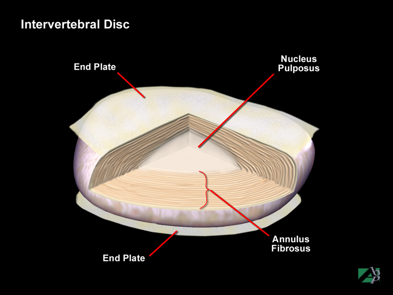
Intervertebral discs are semi-elastic structures interposed between each vertebra. Their purpose is to allow vertebrae to move over each other sideways, forward and backwards and to act as shock absorbers for the spinal column. An intervertebral disc consists of cartilaginous end plates, the annulus fibrosus and the nucleus pulposus. The end plates adhere to the annulus fibrosus and the nucleus pulposus and receive bone marrow from the vertebrae to aid disc nutrition. The annulus fibrosus consists of sheets of collagen fiber that envelops the nucleus and helps prevent structural failure. The elastic annulus is resilient, so that after being stretched it can recoil and springs back to recover after mechanical deformation.
The nucleus pulposus comprises a mass of watery gel that allows it to spread out under pressure preventing it from being compressed. It has a large content of proteoglycans (large molecules comprised of protein and polysaccharides) whose function is to retain water. Intervertebral discs progressively lose their fluid content with age. As the nucleus pulposus loses fluid it is replaced by fibrous tissue and the annulus undergoes degeneration. The fibers making up the annulus then lose their elasticity and the nucleus loses its resiliency
Intervertebral Foramen
An opening between two adjacent vertebrae through which the spinal nerves and blood vessels pass
Intima
The inner layer of an artery
Intra-abdominal
Pertaining or referring to something occurring within the abdomen
Intra-articular
Pertains or refers to being within a joint
Intracapsular
Pertains or refers to being within the capsule of a joint
Intracapsular Fracture
A fracture occurring within the capsule of a joint
Intracerebral Hematoma
Intracerebral hematoma refers to hematoma formation from hemorrhage within the brain matter itself. In most cases intracerebral hemorrhages are multiple and also are often seen with extracerebral hemorrhaging and other concomitant brain trauma. The most common site for intracerebral hematoma is the temporal lobe accounting for about three quarters of intracerebral hematomas. Less common sites are the frontal lobes and the parietal and occipital lobes. The majority of large intracerebral hemorrhages are fatal. Death results from rupture into the ventricular system or by swelling resulting in herniation of the temporal lobes through the tentorium cerebelli. This results in displacement and compression of the midbrain, hemorrhage of the midbrain and pons, and eventually death
Intracranial
Within the skull, the brain or its meninges
Intracranial Hematoma
A hematoma between the meninges of the brain. The meninges comprise three layers, with a potential space between each layer. The inner layer is the pia mater, the middle layer the arachnoid and the outer layer the dura. A hematoma between the pia mater and arachnoid is known as subarachnoid hematoma, a hematoma between the arachnoid and the dura is known as a subdural hematoma and a hematoma between the dura and the skull is called an epidural hematoma
Intracranial Neurostimulator
An intracranial neurostimulator is something like a pacemaker. The Neurostimulator delivers impulses to electrodes implanted within the skull to suppress certain brain activities. It is mainly used to suppress chronic pain and uncontrollable tremors
Intraluminal
This term describes the inside of a hollow structure or organ such as the esophagus or an artery. The term may be used to describe an injury, for example an intraluminal injury to the esophagus, which would mean it was an injury inside the esophagus, such as from a burn or from ingesting some caustic substance
Intramedullary Nail
An internal fixation device used to secure fractures of long bones, particularly the femur. The nail is inserted into the medullary canal (bone marrow)
Intraocular
Within the eyeball
Intrathecal Catheter
An intrathecal catheter or pump implant is a medical device implanted for the relief of chronic intractable pain. The device consists of an infusion pump, a spinal catheter and an external programmer. The infusion pump is placed under the skin in the right or left side of the abdomen in a pouch created in the skin to hold the pump. The catheter is placed through a small incision in the back and is tunnelled from the back underneath the skin to the abdomen where it is connected to the pump. The pump is filled with a pain reliever often morphine
Intravenous
Intravenous (IV) means via or through the veins. Intravenous infusion could involve feeding nutrients, fluids, antibiotics, anticoagulants or other therapeutic substances. Intravenous infusion allows for quicker uptake of substances into the body
Intubation
Intubation is an emergency procedure to provide an adequate airway for resuscitation. It is often the first step in the resuscitation phase for unconscious or semi-conscious trauma victims. There are a number of different intubation devices used but all are primarily tubular devices passed into the airways to keep the airway open and unobstructed
Iridectomy
Removal of part or the entire iris
Iridocyclitis
Inflammation of both the iris and the ciliary body of the eye
Iridotomy
Iridotomy is a procedure where a hole is punched through the iris (the pigmented colored segment of tissue around the pupil) to relieve intraocular (within the eyeball) pressure. This procedure may be done using a laser
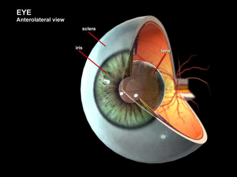
Iris
The circular pigmented part of the eye; the center of the eye is the pupil
Irrigation
To wash out, with water or some other solution
Ischemia
Lack of blood supply to a bodily organ or structure
Ischial Tuberosity
A bony projection on the ischium of the pelvis
Ischium
The lower back part of the pelvis
Islets of Langerhans
Part of the pancreas, they are masses of cells within the pancreas, their function is to produce insulin
Isometric Exercises
A form of physiotherapy in which the contraction of one set of muscles is counteracted by opposing muscles