Medical Glossary - Letter R
This medical glossary of terms beginning with the letter "R" contains the more common medical terms one might expect to encounter in a medical report or in hospital notes. The glossary is intended as a quick reference only; many of the terms are also referenced and illustrated in more detail in the medical libraries, to which you should refer for more detailed information.
Radial
Refers to the radius or the radial side of the forearm, the thumb side of the forearm
Radial Artery
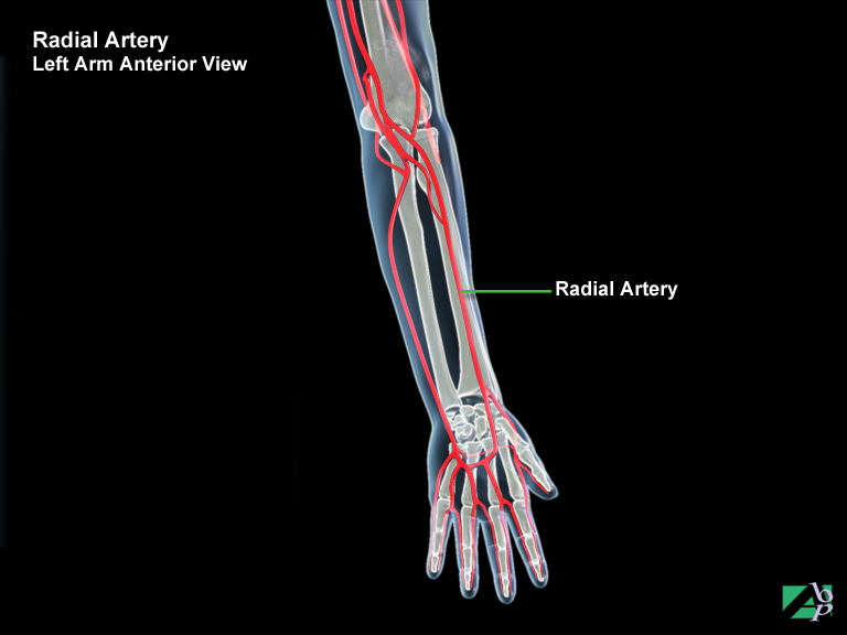
The radial artery is a continuation of a branch of the brachial artery. It descends from just below the elbow and runs down the radial side of the forearm (the thumb side) to the wrist and across the palm where it merges with a branch of the ulnar artery. It supplies blood to the radial side of the forearm
Radial Collateral Ligament
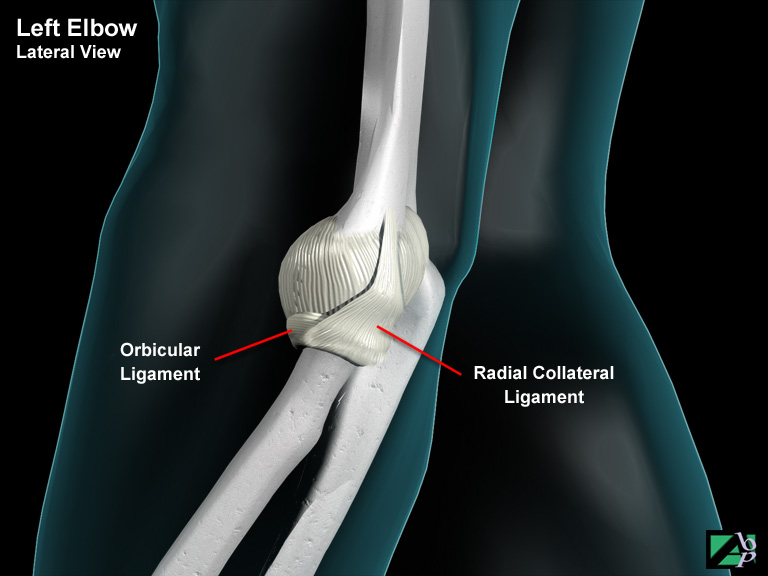
A ligament of the elbow
Radial Keratotomy
Radial keratotomy is a procedure in which a series of fine microscopic incisions are used to flatten the cornea. It is performed to correct refractive errors e.g., short sightedness and long sightedness
Radial Nerve
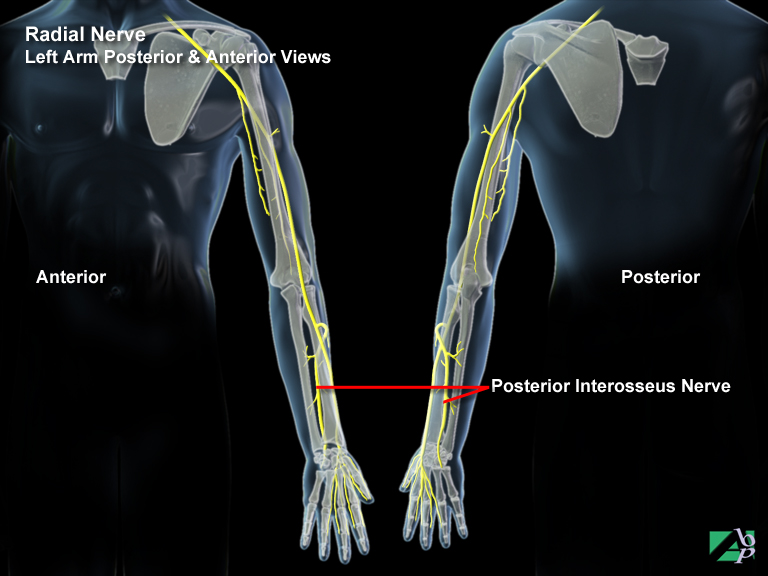
The radial nerve is a mixed nerve but predominantly is a motor nerve. Its sensory branch supplies sensation to the skin of the back of the hand and wrist area and the back of the index finger and thumb. Its motor fibers innervate the extensor muscles of the elbow, forearm, wrist, fingers and thumb
Radial Tunnel Syndrome
Radial Tunnel Syndrome is an entrapment syndrome. There are several places the radial nerve can be compressed at or around the elbow. At the lateral portion of the elbow, the nerve travels in a tunnel that is formed by the surrounding muscles and bone. If the tunnel is too small for any reason, the nerve can be squeezed and begin to cause pain. Repetitive forceful pushing and pulling, bending of the wrist, gripping and pinching further stretch and irritate the nerve. Sometimes a direct blow to the lateral side of the elbow may also injure or damage the radial nerve. Constant use of the arm for twisting activities such as might be found on an assembly line can cause irritation on the radial nerve and also lead to radial tunnel syndrome.
Radical Excision
A radical excision means a wide or extensive or complete excision, for example in the case of a skin lesion, it would mean excision of the lesion and adjacent or underlying tissue in order to completely eradicate the lesion
Radicular
Refers to a spinal nerve root
Radiculitis
Inflammation of a spinal nerve root
Radiocarpal
Pertaining to the radius and the carpal area, or the wrist
Radiograph
An x-ray
Radioulnar
Refers to the radius and the ulna
Radioulnar Synosis
Same as cross union, it describes abnormal bony union between the proximal radius and ulna at the elbow
Radius
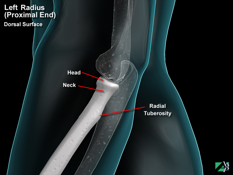
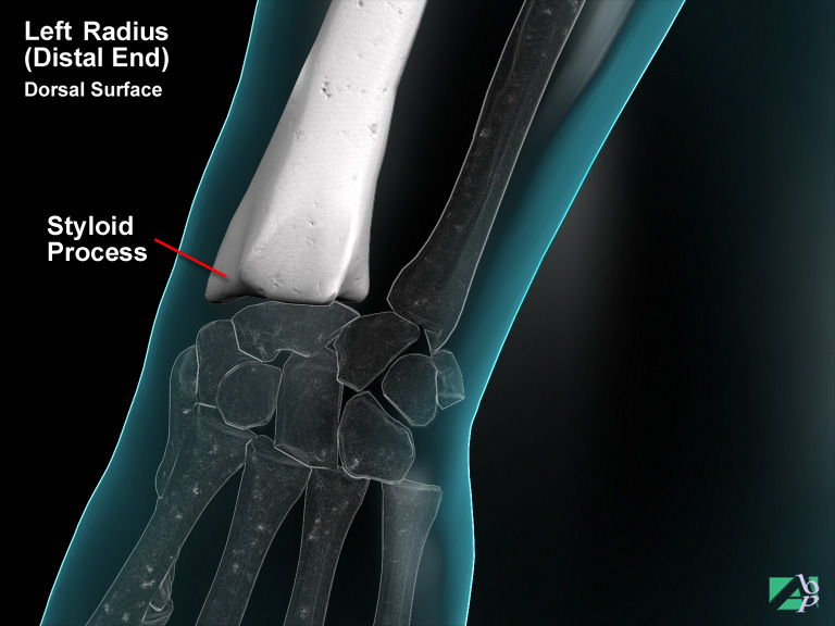
The radius is a bone of the forearm; it comprises a proximal (elbow) end, a shaft and a distal (wrist) end. The proximal radius is the end of the radius nearest the elbow (it plays only a minor role in elbow function). It is comprised of a small disc shaped head that connects with the capitulum of the humerus and the radial notch of the ulna. Below the head is the neck and radial tuberosity. The latter, which is a bony elevation, is where the tendon of the biceps brachii muscle attaches and where it binds with the ulna. The lower end (wrist end) of the radius has a concave notch called the ulna notch, which forms a joint with the head of the ulna. Like the ulna, it also has a styloid process, which provides an attachment point for the tendon of the brachioradialis muscle and for the radial collateral ligament of the wrist joint. The outer surface of the process has a groove for the passage of the abductor pollicis longus, which abducts the thumb and the extensor brevis muscles that extend the hand
Rale
An abnormal sound that might be heard from the chest using a stethoscope
Raynaud's Syndrome (Disease)
This is a neurovascular disorder in which the digits (usually the fingers) undergo a reaction to cold. Blood vessel constriction results, causing pain and changes in skin color. The cause is unknown but it is usually attributed to attacks of severe vasospasm in the digital arteries. The attacks are usually precipitated by exposure to cold but may also be precipitated by emotional distress. The condition is also associated with a number of diseases such as rheumatoid arthritis, systemic lupus, sclerosis and long-term exposure to certain chemicals such as vinyl chloride. A connection may also exist with the use of vibrating tools. Women are affected more than men
Rectus Capitus Anterior
A muscle in the back of the neck originating from the Atlas (C1) and inserting onto the occipital bone of the skull. It assists flexion of the Atlanto-occipital joint
Rectus Capitus Lateralis
A muscle in the back of the neck originating from the Atlas (C1) and attaching to the occipital bone of the skull. It assists lateral flexion of the Atlanto-occipital joint
Rectus Capitus Posterior Major
A muscle in the back of the neck that originates from C2 and attaches to the occipital bone. It assists extension and rotation of the Atlanto-occipital joint
Rectus Capitus Posterior Minor
A muscle in the back of the neck that originates from C1 and inserts onto the occipital bone. It assists extension at the Atlanto-occipital joint
Rectus Femoris
A muscle of the upper leg originating from the iliac spine of the pelvis and attaching to the patella and tibia. It assists extension of the leg at the knee and flexion of the thigh at the hip. The femoral nerve innervates it
Referred Pain
Pain felt in a region of the body where the source of the pain comes from another area of the body. For example sciatica produces pain that radiates down the leg but the origin of the pain may be due to irritation of the sciatic nerve in the lower back, or a liver disorder may produce shoulder pain
Reflex Sympathetic Dystrophy
This syndrome describes a set of symptoms that include pain, and trophic skin changes (blotchy, shiny skin, warm to the touch), and because of pain and disuse of the affected body part, muscle wasting, joint stiffness and fixed deformities may also be seen. Numerous clinical conditions are covered under the syndrome including hand-shoulder syndrome and Sudeck's atrophy. More recently it has been referred to as "Complex Regional Pain Syndrome". Despite a plethora of theories, the cause of the syndrome is not known; some consider it is due to a disorder of the sympathetic nervous system, others consider the main cause to be psychosomatic. It may be precipitated by trauma, even trivial, but is more often seen with injuries such as fractures and mostly associated with injuries to the hand or foot
Removable Bridge
A removable bridge is a partial denture. A false tooth or teeth are held in place by clips or clasps. The bridge can be removed by the patient, as opposed to a fixed bridge, which cannot
Renal
Referring to the kidneys
Renal Artery
The renal artery is a branch of the abdominal aorta that supplies blood to the kidney
Reticular Activating System
A complex network of nuclei and nerve fibers that function to arouse the cerebrum. Portions of this network are located in the spinal cord, pons and midbrain, thalamus and hypothalamus. The principle functions of the reticular activating system are to keep the cerebrum in a state of alert consciousness and to selectively monitor sensory impulses perceived by the cerebrum. The reticular activating system also aids the cerebrum to maintain contraction of skeletal muscles
Retina
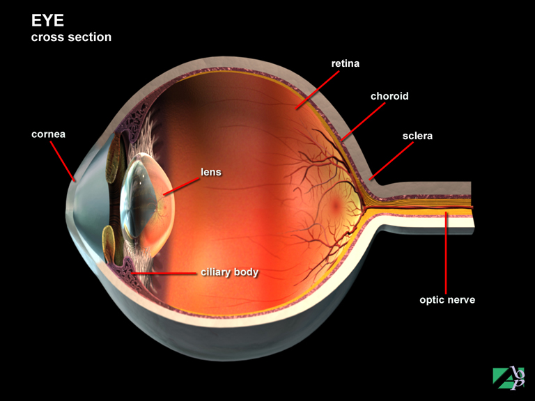
The retina, which covers the choroid, is the inner neural layer of the eye. It comprises two portions, an outer-pigmented layer and a neural layer. The two layers are not attached to each other except at two points. Because of this loose connection the retina may become detached. In the center of the outer layer is the macula, which is vital for clear vision. The all-important neural layer is composed of many varied nerve cells and photoreceptors (cone- and rod-shaped components sensitive to light stimulation). The information gathered by the photoreceptors is integrated into a visual message conveyed by the optic nerve to the brain. Rods are located in the peripheral parts of the retina and respond to dim light for black and white vision. They also respond to shape and movement but do not provide good visual acuity. The cones are located near the center of the retina (the macula) and respond to daylight color vision and are responsible for visual acuity
Retinal
Relating to the retina of the eye
Retrobulbar
Retrobulbar refers to the space in the eye socket behind the eye
Retrograde Amnesia
Loss of memory of events leading up to a head injury
Retroperitoneal
This means behind the peritoneum, which is the membrane that covers the lining of the abdominal wall
Retroperitoneal Fistulogram
Retroperitoneal refers to behind the peritoneum, which is the membrane that lines the interior surface of the abdomen. A fistula is an abnormal opening. A fistulogram involves injecting a radiopaque dye into the fistula to better visualize the fistula for x-rays to be taken
Rhinitis
Inflammation of the mucous membrane of the nose
Rhinoplasty
Rhinoplasty is a plastic procedure generally performed to improve the appearance of the nose
Rhinorrhea
The term rhinorrhea refers to a watery discharge from the nose (as seen in colds). CSF rhinorrhea refers to leakage of cerebrospinal fluid from the nose
Rhizotomy
A rhizotomy is the surgical cutting or division of a nerve root; in trauma cases it is performed most frequently on spinal nerves for pain relief in cases of intractable pain. It may also be performed on cranial nerves for individuals with intractable facial neuralgia
Rhomboid Major
A muscle of the back originating from the spinous processes of T2-T5 and inserting onto the scapula. It retracts and rotates the scapula. It is innervated by the dorsal scapula nerve
Rhomboid Minor
A muscle of the upper back originating from the spinous processes of C7 and T1 and inserting onto the scapula. It assists retraction and rotation of the scapula. The dorsal scapula nerve innervates it
Rib
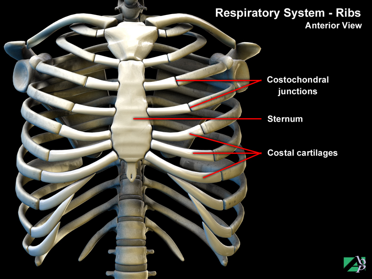
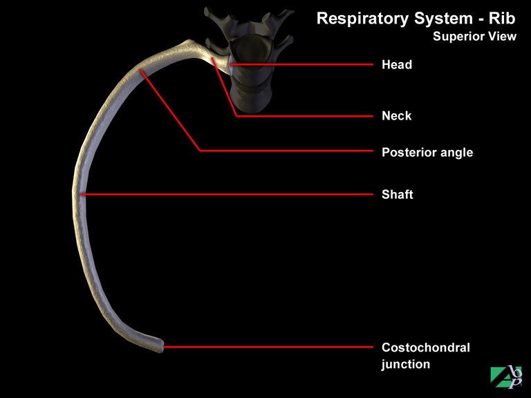
There are 24 ribs, or 12 pairs of ribs. Posteriorly (at the rear) each pair attaches to a thoracic vertebra. Anteriorly (in front) the first seven pairs attach to the sternum through connections with the costal cartilages, which attach to both the ribs and the sternum. The paired ribs 8, 9 and 10 are attached to the costal cartilage of the seventh rib. Ribs 11 and 12 do not have any anterior attachment and are often referred to as floating ribs. With the exception of ribs 11 and 12, each rib has a head, tubercle, neck, angle and a shaft. The head and tubercle are for articulations with the body and transverse processes of the thoracic vertebra. Because ribs 11 and 12 do not attach to the vertebra they are missing these anatomical parts. The neck lies just behind the head. The shaft or body is the main curved part of the rib. The angle is the point where the curve is most pronounced. The space between each rib is called the intercostal space where the intercostal muscles are located
Rib Cage
This refers to the bony framework that comprises the 12-paired ribs, the sternum and the 12 thoracic vertebrae of the spine
Right Hypochondriac Area of Abdomen
The right hypochondriac region contains the gallbladder, portions of the liver and the right kidney. It describes the location of the right upper one third of the abdomen
Right Inguinal Region of Abdomen
The right inguinal region contains the appendix, some of the cecum and the small intestine. It describes the location of the right lower one third of the abdomen
Right Lateral Region of Abdomen
The right lateral region contains the cecum, ascending colon, portions of the right kidney and the small intestine. It describes the location of the right lateral one third of the abdomen
Risorius
A facial muscle on the side of the face. It acts to draw the angle of the mouth laterally
Root Canal Therapy
The root canals are structures that house the pulp of a tooth, which is made up of tiny blood vessels and nerves. In root canal therapy, the pulp is removed through a small opening made in the crown of the tooth. Tiny files are then passed down through the opening into the canals to clean and disinfect them. The canals are then sealed with inert material to fill in the gap made by the removal of the pulp. Root canal therapy is usually performed before applying a crown. Root canal therapy is usually performed over a number of visits
Rotator Cuff
The rotator cuff is the name for a group of muscles; it is comprised of four muscles: the supraspinatus, infraspinatus, teres minor and subscapularis. These four muscles arise from the scapula (the shoulder blade) and their tendons blend with the joint capsule to form the rotator cuff