Medical Glossary - Letter K
This medical glossary of terms beginning with the letter "K" contains the more common medical terms one might expect to encounter in a medical report or in hospital notes. The glossary is intended as a quick reference only; many of the terms are also referenced and illustrated in more detail in the medical libraries, to which you should refer for more detailed information.
Keloid
Raised unsightly skin forming on wound, a term often used to describe the state and extent of a scar
Keratitis
Inflammation of the cornea
Keratomileusis
Keratomileusis is an operation on the cornea performed for refractive errors in which a layer of the cornea is removed, re-shaped and then re-attached to the cornea
Keratophakia
Keratophakia is a form of keratoplasty (corneal transplant) in which corneal tissue from a donor is frozen, shaped and inserted into the cornea of the patient
Keratoprosthesis
Keratoprosthesis is a plastic replacement for an opacified part of the inner cornea usually performed for cataract; it may be a temporary procedure
Kidneys
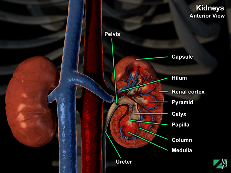
The kidneys are the functional unit of the urinary system, which comprises the kidneys, the paired ureter, the urinary bladder and the urethra. The two bean shaped kidneys lie on each side of the vertebral column at a level from the 12th thoracic vertebra to the 3rd lumbar vertebra. They are described as being retroperitoneal, which means they are outside the peritoneum and therefore not strictly within the abdominal cavity. The right kidney is usually lower then the left because of the position of the liver above. Each kidney consists of three layers. The innermost layer is the renal medulla, the second layer is called the adipose capsule and the outer layer is known as the renal cortex, which is surrounded by a fibrous structure called the renal capsule. The kidneys are anchored to the abdominal wall by the peritoneum. The function of the kidneys is to extract urine from the liquid portion of the blood and the waste materials dissolved in it. Blood is circulated continuously through the kidneys and units within the kidney called nephrons do the filtering. There is estimated to be over one million nephrons within each kidney. A nephron consists of urinary tubules and small blood vessels. Each filtering unit comprises a renal corpuscle (a tuft of blood capillaries) and a long slender tube called a tubercle, which is a collecting duct for the urine. Blood passes through the walls of the renal corpuscle and into the tubercle where the urine is formed and collected by cup shaped structures called calyces and then passed out of the kidneys into the ureter and then on to the urinary bladder
Kienbock's Disease
A disease of the lunate bone in the wrist. It is caused by inadequate blood supply to the bone and results in avascular necrosis of the lunate
Kirschner Wires
Wires used to hold (stabilize) a reduced fracture; they may be used for example to secure reduced fractures of the patella or facial fractures. Sometimes referred to as just "K" wires
Knee
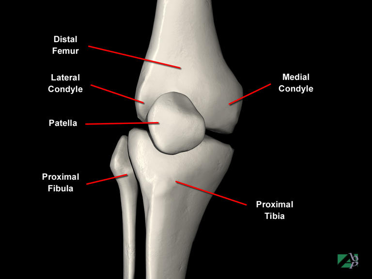
The knee joint is formed by the connection (articulation) between the femur and the tibia. The medial and lateral condyles of the distal femur connect with an expanded area of the tibia called the lateral and medial plateau. The medial condyle of the femur sits on the medial tibial plateau and the lateral femoral condyle sits atop the lateral tibial plateau. In front of the femoral condyles, lies the patella (kneecap). The patella, or as it is more commonly known, the kneecap, is a rounded triangular shaped bone, which serves to protect the knee joint and to strengthen the quadriceps tendon. It is a sesamoid type of bone, which means that it is a bone that is situated within a tendon, in this case the quadriceps tendon. At birth, the patella is actually formed entirely of cartilage, which later ossifies (converts) into bone. The patella connects with the femoral condyles and glides up and down a trough or track on the femur; it constantly changes position with knee movements
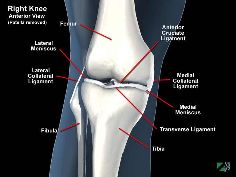
Because of its weight bearing and functionality it provides for human mobility, the knee joint capsule is a complex arrangement of ligaments, tendons, cartilage and other soft tissue. The two menisci of the knee, the lateral and medial meniscus are crescent shaped cartilages that lie between the femur and the tibia. They assist load bearing, act as shock absorbers and lubricate the joint. The knee joint is bound together by numerous ligaments: the ligamentum patellae, the medial and lateral collateral ligaments, popliteal ligaments and the anterior and posterior cruciate ligaments.
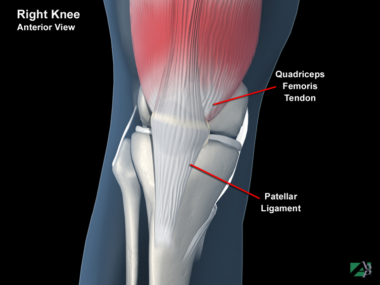
Powerful muscles and tendons including the quadriceps muscles and tendon, patella ligament and tendon, patellotibial tendon and the gastrocnemius muscles provide knee movements. As with any joint in the body, the knee contains several bursae whose main function is to minimize the friction between the femur and the tibia and the patella. A bursa is a sac filled with synovial fluid. Synovial fluid provides lubrication for the joint
Knuckle
The prominence formed at the metacarpophalangeal joints when a fist is formed, the metacarpophalangeal joint is the joint formed by the base of the phalanges (fingers) and the heads of the metatarsals
Kyphoscoliosis
This term describes abnormalities of the normal curvature of the spine. The term derives from kyphosis and scoliosis, which are two separate spinal curvature abnormalities. Kyphosis describes a humped spine; scoliosis is an abnormal side-to-side curvature of the spine. Kyphoscoliosis therefore describes both a humped deformity and an abnormal side-to-side curvature of the spine
Kyphosis
An abnormal curvature of the spine resulting in the spine being humped, also known as hunchback