Medical Glossary - Letter C
This medical glossary of terms beginning with the letter "C" contains the more common medical terms one might expect to encounter in a medical report or in hospital notes. The glossary is intended as a quick reference only; many of the terms are also referenced and illustrated in more detail in the medical libraries, to which you should refer for more detailed information.
Caecum
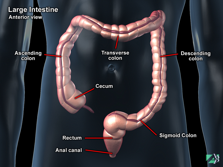
The first part of the large intestine. It is a dilated pouch that hangs behind a membrane known as the ileocecal valve, which serves to prevent the backflow of digested material into the stomach
Calcaneal Spur
A bony outgrowth on the calcaneus, the heel bone
Calcaneal Tuberosity
An extension on the back of the calcaneus (heel bone), which serves as a point of attachment for calf muscles and the Achilles tendon
Calcaneofibular Ligament
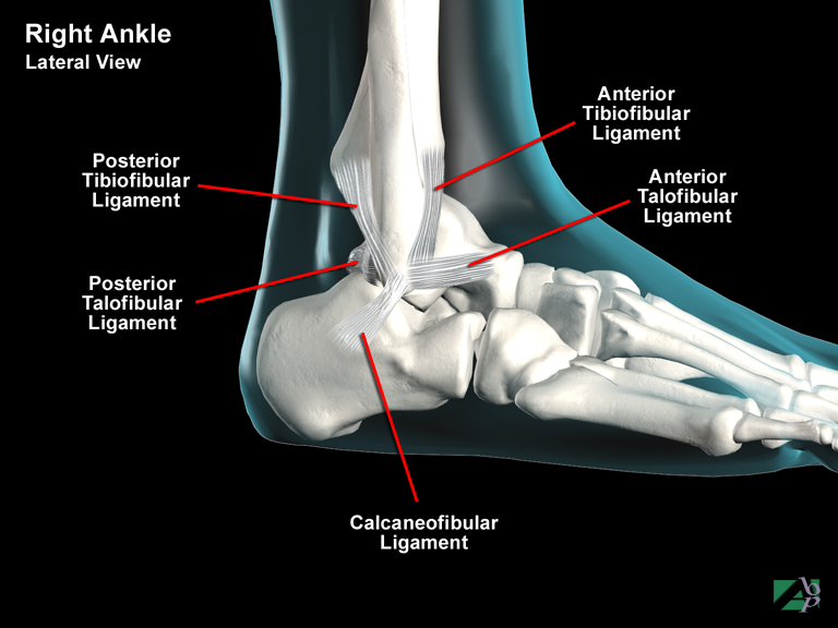
A ligament in the ankle that helps support the ankle joint
Calcaneonavicular
Relating to the calcaneus (heel bone) and the navicular, one of the bones of the hind foot
Calcaneus
The heel bone, the largest of the talar bones. It lies below the talus (which it supports) and behind the navicular and cuboid bones. A posterior extension of the bone called the calcaneal tuberosity is for the attachment of the calf muscles and the Achilles tendon
Calcify
To deposit calcium, to harden. A term often used to describe the progress of bony union following a fracture
Calcifying Tendinitis
Calcifying tendinitis is inflammation of a tendon caused by deposits of calcium. Deposits of calcium can infiltrate any tendon, ligament or joint capsule but the shoulder and the rotator cuff is by far the most common site. The cause for the calcium deposits isn't known but trauma can precipitate calcifying tendinitis. Other associations are hypoparathyroid disease with high blood calcium levels and in people who drink a lot of milk. Calcium deposits are seen more often in women than men and may also be seen occasionally in children
Calculus
A stone like mass as in kidney stones or gallstones, it is formed from mineral salts
Callus
This term may be used to describe new bone formation formed following trauma to a bone, or it may also be used to describe an area of skin, which has become thickened and hardened, as from manual labor
Calyces
Cup shaped receptacles in the kidneys where urine collects
Canaloplasty
A plastic repair procedure to the external auditory canal of the ear
Cancellous Bone
Cancellous bone is bone with a sponge like appearance. The inner surfaces of the cranial bones are made of cancellous bone whereas the outer surfaces are made of what is called compact bone
Canthoplasty
A procedure on the eyelid to correct its position
Canthorrhaphy
A surgical procedure to an eyelid to correct a deformity
Canthotomy
Canthotomy is the surgical incision of the canthus, which is the angle formed by the joining of the upper and lower eyelids on either side of the eye
Canthus
The corners of the eye formed by the junction of the upper eyelid and lower eyelid. The one nearest the nose is called the medial canthus and the one nearest the cheek is the lateral canthus
Capillaries
Tiny blood vessels found throughout the body, they connect arteries to veins
Capitate
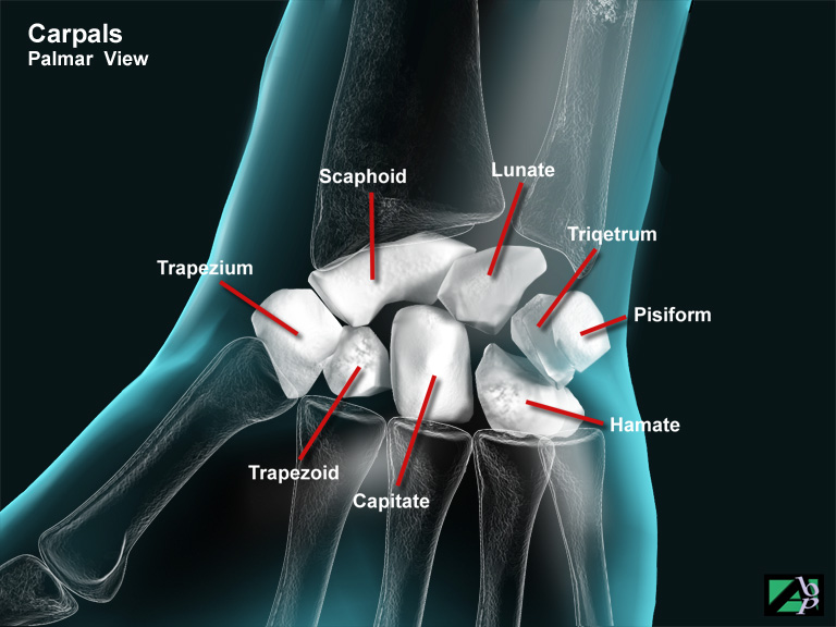
One of the bones of the wrist. The capitate is the largest of the carpal bones and has connections with the hamate and trapezoid on either side of it, the scaphoid and lunate behind and with the 3rd and 4th metacarpals of the hand
Capitellum
A small rounded protuberance on a bone such as on the distal humerus (elbow)
Capsulectomy
A procedure to remove the secondary membrane that holds the lens in place after previous cataract removal surgery
Cardiac Retraining
An outpatient program of graded and monitored exercises designed to gradually increase cardiac capacity. A cardiologist may recommend that an individual participate in this type of program following any injury or surgery to the heart
Cardiovascular Stress Test
This is a test performed by having an individual walking or running on a treadmill for a specified period of time while being monitored by a cardiologist or a designee. Monitoring can include blood pressure, pulse, heart rate and EKG recording. The purpose of the test is to determine the heart's ability to function under stressful conditions
Carpal Bones
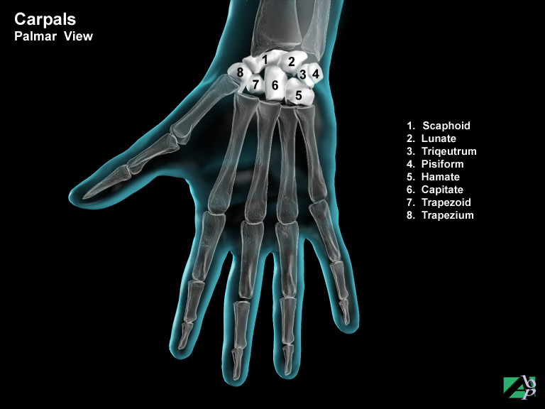
This term refers to the wrist bones, there are eight in all, in two rows, comprising a proximal row and a distal row, they are: the scaphoid, lunate, triquetrum, pisiform, hamate, capitate, trapezium and trapezoid. The proximal row refers to those bones nearest the forearm; the proximal row comprises the scaphoid, lunate, triquetrum (also known as the triangular) and the pisiform. The distal row, the row nearest the hand is comprised of the hamate, capitate, trapezium and the trapezoid
Carpal Tunnel
This refers to a space between the carpal bones and the carpal ligament, through which the median nerve and extensor tendons of the hand passes. It is located deep within the wrist on the palmar side i.e., on the same side of the wrist as the palm of the hand
Carpal Tunnel Syndrome
The term carpal tunnel syndrome describes a set of symptoms arising from compression of the median nerve at the wrist. The median nerve, along with the tendons that flex the fingers pass together through a space in the wrist known as the carpal tunnel. This tunnel is narrow and even in the normal individual barely has room to accommodate the nerve and the tendons. Any inflammation or irritation of these structures causes compression of the median nerve. The causes of carpal tunnel syndrome are controversial, repetitive wrist movements have been blamed but in many cases the condition occurs without any plausible reason. Other known associations are menopause, pregnancy and trauma, in the latter case compression from fractures and dislocations. It is much more likely to affect females than males and in many cases is bilateral. The symptoms of the syndrome are pain and paresthesia in the palm of the hand, thumb index and middle fingers
Carpometacarpal
This refers to the area where the carpal (wrist bones) and metacarpal (hand bones) meet
Carpometacarpal Joint
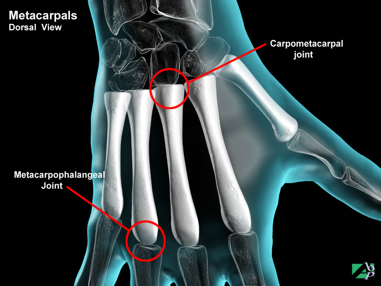
The joint (articulation) between a carpal bone (wrist bone) and a metacarpal (hand) bone
Carpus
The carpus or bones of the wrist consists of eight bones called carpal bones, which are arranged into two transverse rows (proximal and distal rows) consisting of four bones per row. The proximal row (the row nearest the distal ends of the radius and ulna) from the thumb side of the hand to the little finger is the scaphoid (also known as the navicular), lunate, triquetrum (also known as the triangular) and the pisiform. The distal row (the row nearest the metatarsals of the hand) from the thumb side of the hand to the little finger is the trapezium, (also known as the greater multangular), trapezoid (also known as the lesser multangular), capitate and hamate
Cataract
An opacity of the retina of the eye
Cathartic
A strong laxative
Catheter
A slender rubber tube used to extract fluid from the body. It is generally used for the bladder and urethra
Cauda Equina
A bundle of spinal nerves that emerge from the spinal cord near the coccyx
Causalgia
Causalgia is a term used to describe a burning like pain sensation. The cause is not clear, although it is thought to be due to damage to sensory nerves supplying the body part producing the sensory changes. Causalgia bears some resemblance to reflex sympathetic dystrophy. Trauma is usually the precipitating factor. Causalgia usually affects the hands or feet
Cecectomy
Cecectomy is the surgical removal or partial removal of the caecum, which is the first portion of the large intestine. This procedure may also involve resection of the terminal portion of the ilium of the small intestine
Celiac Artery
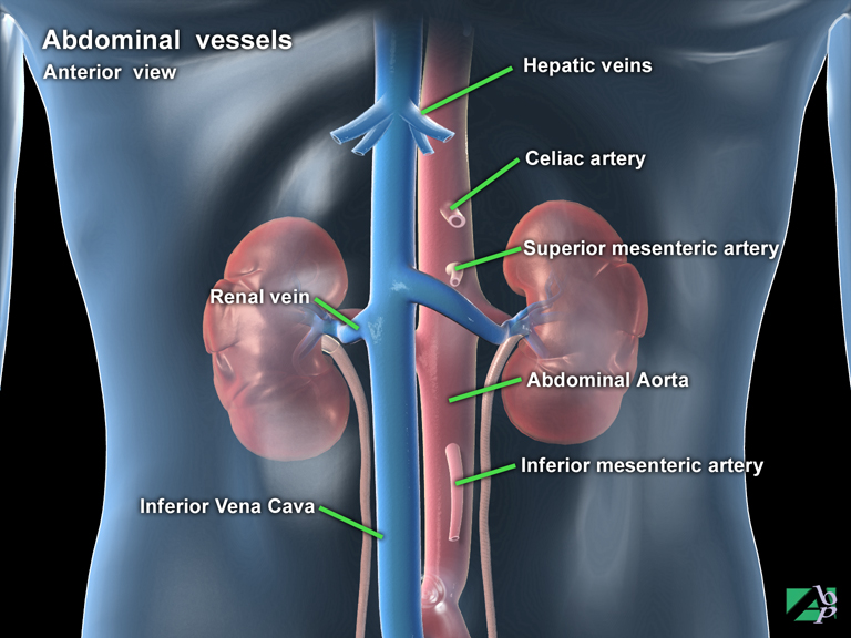
The celiac artery, which is a branch of the abdominal aorta supplies blood to the esophagus, stomach and part of the small intestine, liver, spleen and pancreas
Central Nervous System
The central nervous system or CNS comprises the brain and the spinal cord
Central Serous Retinopathy
Central serous retinopathy, also known as chorioretinopathy, is a collection of fluid under the retina that results in visual distortion. Symptoms include a blind spot, decreased or blurry version and distortion of shapes. In many cases the condition resolves spontaneously within about 6 months although many suffer recurrences
Cerclage
A medical procedure where a structure is tied together with a wire loop or ring
Cerebellum
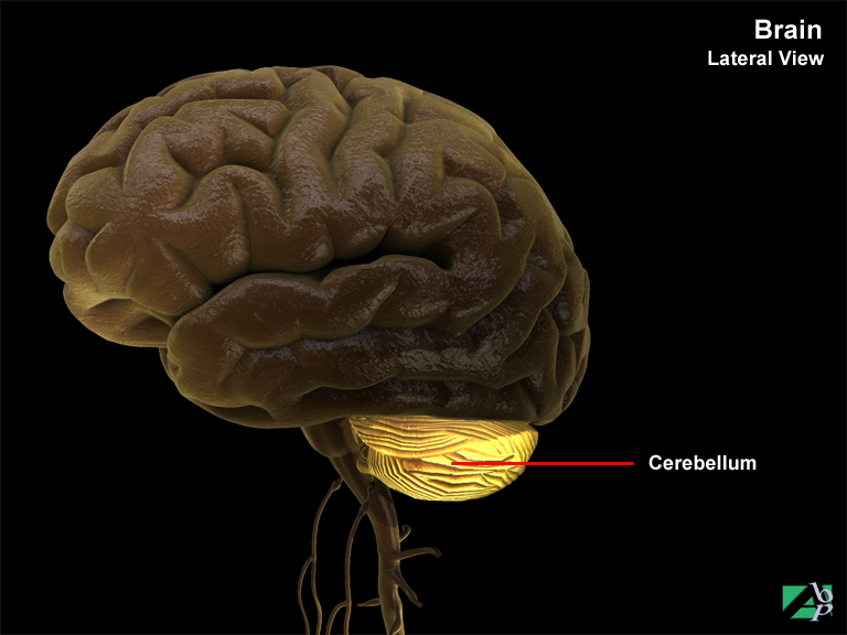
The cerebellum (or as sometimes referred to as the little brain) is the second largest structure of the brain and occupies the inferior and posterior aspects of the cranial (skull) cavity. The cerebellum consists of two hemispheres and a central constricted area called the vermis. Like the cerebrum, the cerebellum has a thin outer layer of grey matter, called the cerebellar cortex and a thick deeper layer of white matter. Three paired bundles of nerve fibers called cerebellar peduncles support the cerebellum and provide it with tracts for communicating with the rest of the brain. The principle function of the cerebellum is coordinating skeletal muscle contractions. Impulses for voluntary muscle movement originate in the cerebral cortex and are coordinated by the cerebellum. The cerebellum constantly initiates impulses to selective motor units for maintaining posture and muscle tone
Cerebral Contusion
Bruising of the brain tissue
Cerebral Cortex
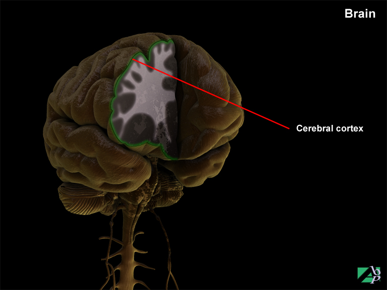
The cerebral cortex is the surface layer of the cerebrum. It is about 1/2 inch in thickness, although this varies slightly in different areas of the brain. It is composed of what is referred to as grey matter and white matter. The grey matter of the cerebral cortex is a densely packed region comprising neurons, dendrites and capillary blood vessels. The neurons (nerve cells) are the basic operative units of the brain. Below the grey matter is a thicker inner layer, the sub-cortical layer, which is white in appearance. The white matter is made up of dendrites and myelinated axons. These neural fibers form the billions of connections within the brain by which information in the form of electrical impulses is transmitted to the selective areas of the brain. There are three types of fiber tracts within the white matter: association fibers, commissural fibers and projection fibers. Association fibers belong to a given hemisphere and only conduct neural impulses within their given hemisphere. Commissural fibers connect neurons from one hemisphere to another. Projection fibers form tracts that transmit and receive impulses from neurons in the cerebrum to other parts of the brain and spinal cord
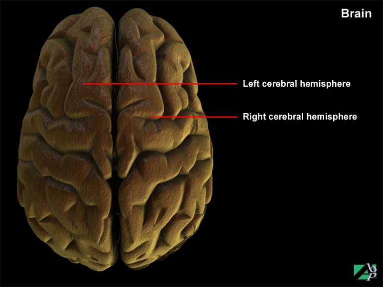
Cerebral Hemispheres
There are two cerebral hemispheres of the brain, named the left and right hemispheres, which are divided into large subdivisions called lobes. Four of the lobes are on the surface of the hemispheres and are named according to the overlying cranial (skull) bones. These lobes are the frontal lobe, parietal lobe, temporal lobe and occipital lobe. A fifth lobe, the insula lies deep in the cerebrum and is covered by portions of the frontal, parietal and temporal lobes. The two hemispheres carry out different functions. Generally the left hemisphere carries out analytical and verbal functions. The right hemisphere is the source for spatial and artistic skills. The hemispheres via the connection with the corpus callosum (white matter) share memory and learning.
Cerebral Laceration
A cerebral laceration is tearing of the brain tissue. It is usually seen with penetrating injuries or is associated with skull fractures where a fracture fragment becomes depressed into the brain tissue
Cerebral Edema
Cerebral edema is swelling of the brain due to an increase of water content. Normal intracranial volume pressure comes from 80 percent brain tissue, 10 percent cerebrospinal fluid and 10 percent from blood within blood vessels. Raised intracranial pressure results when there is an increase in volume in one of these three without a compensatory decrease in the volume in one of the other two.
Cerebral Ventricles
Cerebral ventricles are cavities or chambers within the brain. The ventricles of the brain are where the cerebrospinal fluid is produced. The fluid is formed by tissue known as the choroid plexus that exists in each ventricle. There are four ventricles, the lateral ventricles are found in the right and left cerebral hemispheres of the cerebrum. The third ventricle is situated between the thalamus and the hypothalamus. The fourth ventricle is situated between the pons in front and the cerebellum in the back
Cerebrospinal Fluid
Cerebrospinal fluid is a clear colorless fluid consisting of glucose, urea, proteins and salts in addition to some white blood cells. Cerebrospinal fluid circulates continuously around the brain where it acts as a cushion against jarring or sudden shocks. It also circulates nutritive substances filtered from the blood. The formation of cerebrospinal fluid is continuous and the direction in which it circulates is from top to bottom i.e., from the lateral ventricles down through the third and fourth ventricles and into basal cisterns. The basal cisterns area dilated portion of the subarachnoid space at the base of the brain. The cerebrospinal fluid is eventually absorbed back into the general venous circulation by way of the arachnoid villi that project into the various venous sinuses of the brain. This formation and absorption rate results in a fairly constant volume of circulating fluid. If there is increased production, decreased absorption or blockage of flow, an increased accumulation of fluid within the ventricles will result, resulting in raised intracranial pressure
Cerebrum
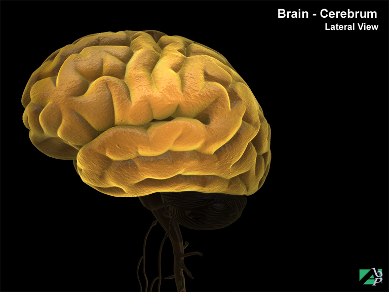
The cerebrum accounts for about 80 percent of total mass of the brain. It consists of the right and left cerebral hemispheres that are divided (though not completely) by a longitudinal fissure. Internally, portions of the two hemispheres are connected by large tract of white matter called the corpus callosum. Each hemisphere fits into the dome of the cranial vault of the skull. The cerebrum comprises a surface layer referred to as the cerebral cortex that is composed of grey matter and below this, a thick layer of white matter. The cerebral cortex features numerous folds and grooves that are called convolutions which form during fetal development. These convolutions allow for a larger area of grey matter that is made up of nerve cell bodies. The elevated fold of the convolutions are called gyri and the depressed groves are called sulci
Cervical
Pertaining to the neck area
Cervical Plexus
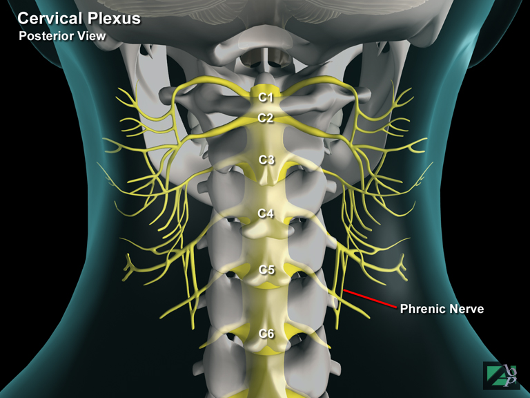
The cervical plexus is formed by the rami of the first four cervical spinal nerves (C1-C4). Most of the nerves arising from this plexus are sensory nerves and supply sensation to the skin of the back of the head, ear, neck and shoulder and upper chest. Two motor nerves also arise from this plexus, the axillary nerve and the phrenic nerve. The axillary nerve innervates the shoulder and the upper arm, while the crucial phrenic nerve innervates the diaphragm, which is essential for breathing
Cervical Rib
An abnormal growth from a cervical vertebra
Cervical Rib Syndrome
A term used to describe neurovascular symptoms in one or both arms produced by compression of the brachial plexus and subclavian artery by a cervical rib, which is an abnormal accessory rib in the neck
Cervicobrachial
Relates to the neck and arm
Chalazion
A chalazion is a small cyst of the eyelid. It is usually caused by distension of the sebaceous gland of the lid, which is a lubricating producing gland. An accumulation of the lubricating material causes the cyst to form
Chemonucleolysis
A medical procedure in which the nucleus pulposus of an intervertebral disc is dissolved with an injection of a chemical substance
Cholangiogram
A cholangiogram is an x-ray of the hepatic bile ducts; it is taken after the injection or introduction of an opaque substance into the ducts
Cholecystectomy
Cholecystectomy is the surgical removal of the gallbladder. The gallbladder is a pear shaped sac that is attached to the underside of the right lobe of the liver. Cholecystectomy may be performed laparoscopically or by open surgery
Cholecystogram
A cholecystogram is an x-ray of the gallbladder. It is taken after the ingestion of an opaque substance
Chondromalacia
Softening of an articular cartilage. Most often it affects the patella cartilage, where it is known as chondromalacia patella
Chondromalacia Patella
Softening of the patella cartilage
Chondrophyte
An abnormality of a cartilage, usually of its growth
Choroid
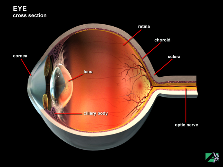
One of the layers of the eye, it lies between the retina and the sclera. It is a vascular layer of the eye and serves to nourish the lens and retina
Choroid Plexectomy
Plexectomy refers to the surgical excision of a plexus, which is a network of interlocking nerves or small blood vessels. The choroid plexus, which consists mainly of blood vessels, protrudes into the ventricles of the brain. Plexectomy is therefore the surgical excision of a choroid plexus from one of the ventricles of the brain
Chorioretinopathy
Same as central serous retinopathy, it is a collection of fluid under the retina that results in visual distortion. Symptoms include a blind spot, decreased or blurry version and distortion of shapes. In many cases the condition resolves spontaneously within about 6 months although many suffer recurrences
Ciliary Body
The ciliary body lies adjacent and is attached to the sclera. One of its basic functions is to nourish the retina. Another is that the muscles of the ciliary body allow the lens of the eye, which is in the center of the ciliary body, to change shape facilitating near and distant vision. The ciliary body also acts as a partition dividing the interior eye into two chambers called the anterior and posterior chambers. These chambers are filled with fluid called aqueous humor, which maintain the intraocular pressure of the eye
Cisternal Puncture
Cisternal puncture is a procedure performed to take a sample of cerebrospinal fluid. A hollow needle is inserted into the cisterna magna, which is a space between the cerebellum and the medulla oblongata
Clavicle
The clavicle or collarbone is a slender S shaped bone that binds the shoulder to the trunk. The middle portion of the bone is tubular while the distal (shoulder) end is flattened in shape. The medial end of the bone (nearest the center of the chest) forms the sternoclavicular joint with the manubrium (upper end of the sternum). The distal end connects with the acromion process of the scapula to form the acromioclavicular joint
Clavicular Notch
A depression in the sternum for articulation with the clavicle
Claw Toe Deformity
Claw toe deformity results from hyperextension of the metatarsophalangeal and flexion of the distal interphalangeal joint. It is caused by contracture of the intrinsic muscles of the foot
Closed Fracture
A fracture not involving exposure of the fractured bone to the outside environment, i.e., with no associated protrusion of the bone or wound at the fracture site
Closed Reduction
A method of reducing a fracture of a bone, or dislocation of a joint by manually manipulating the broken ends or bones back into their normal anatomical position
Coagulate
To form a clot
Coccygectomy
Excision of the coccyx
Coccyx
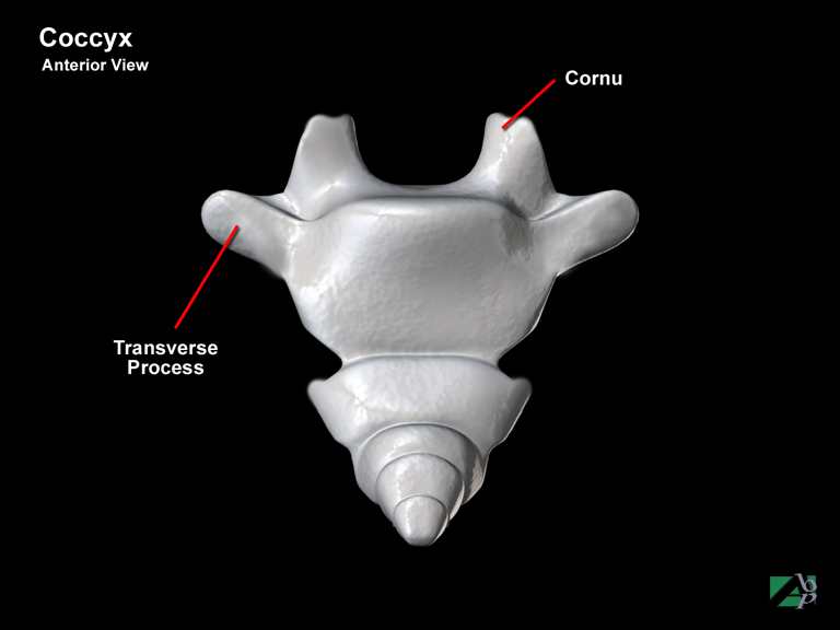
The coccyx is commonly referred to as the tailbone. It is a triangular shaped bone lying below the sacrum and is attached to the sacrum by ligaments. It differs from other spinal vertebra in that it does not contain a central canal. Four important muscles attach to the coccyx, the gluteus maximus, the coccygeus, the sphincter ani and the levator ani
Cochlea
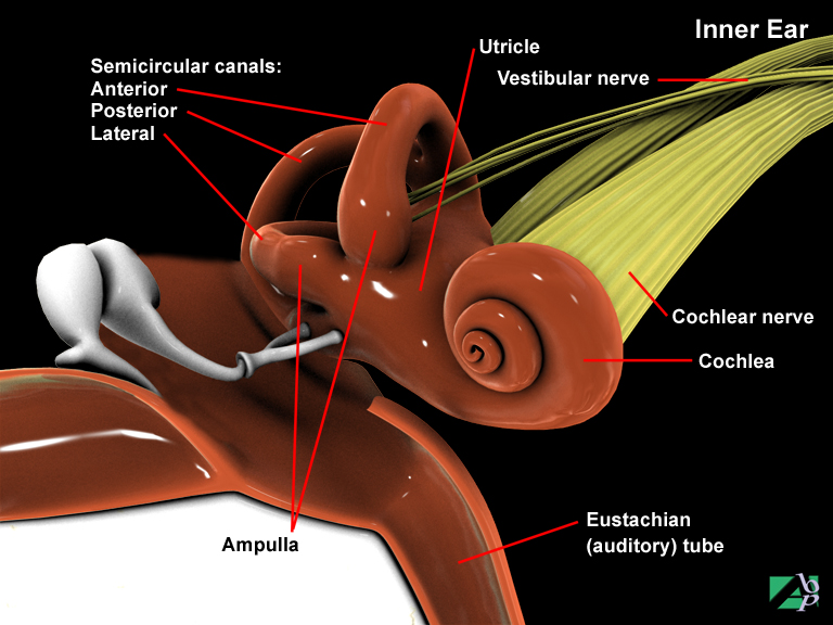
Part of the inner ear situated in the temporal bone of the skull
Colic
Acute abdominal pain
Colles Fracture
A fracture of the wrist, at the distal end of the radius
Colon
The major part of the large intestine. It is divided into three sections named after the direction it travels, e.g., ascending colon, transverse colon and descending colon
Colostomy
A colostomy is the formation of an artificial outlet (a stoma) for the diversion of the contents of the colon to pass through the abdominal wall. An opening is made into the colon, which is then sewed to an opening made in the abdominal wall through which the fecal matter is discharged into a bag called a colostomy bag, which is worn over the abdominal opening. This procedure may be performed because of obstruction of the large bowel or after surgery to the colon. Colostomy may be permanent or temporary. A temporary colostomy may be required until a repaired section of the bowel heals
Comminuted Fracture
A fracture in which the bone is split into several fragments
Common Bile Duct
A duct that empties bile from the liver and gallbladder into the duodenum, the bile assists in breaking down food
Common Carotid Artery
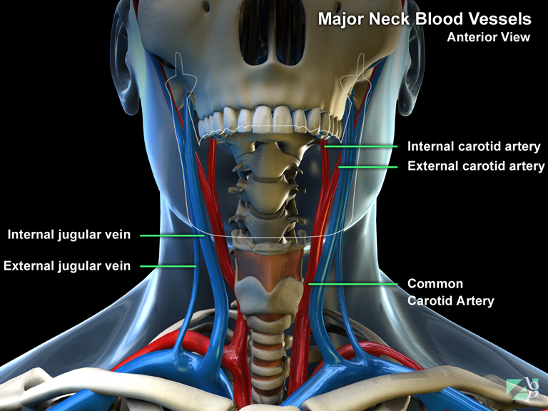
There are two common carotid arteries, one on each side of the neck. As they ascend the neck they divide into an external carotid and internal carotid on each side of the neck. On the left side of the neck, the common carotid arises from the aorta. On the right side it arises from the innominate artery
Compact Bone
Dense hard bone, the long skeletal bones are comprised of compact bone
Compartment Syndrome
A serious limb threatening condition in which blood vessels are compressed within a confined space. This leads to vascular insufficiency to the area dependant on the blood supply from the compressed vessel(s). The tibial compartment of the lower leg is the compartment most affected
Complete Fracture
A fracture where the fracture line extends completely through the bone
Compound Fracture
A fracture in which an end of the fractured bone protrudes through the skin
Compression Fracture
A fracture resulting from compression forces, the thoracic vertebrae are most vulnerable to fractures from compression forces
Computerized Tomography (CT or CAT Scan)
CAT (Computerized axial tomography) or CT (computed tomography) uses special x-ray equipment to obtain image data from different angles around the body, then uses computer processing of the information to show a cross-section of body tissues including organs
Concussion
This describes a condition caused by a severe blow to the head or a fall. The term is generally used to describe a temporary altered mental status without lasting physical effects. It is characterized by symptoms such as dizziness, loss of consciousness, slow breathing, and weakened pulse, vomiting, pallor and fall in body temperature. There may be visual disturbances and confusion. Concussion may also be accompanied by amnesia about events immediately before and after the head injury. Amnesia is described as being retrograde and anterograde. Retrograde amnesia describes loss of memory for events leading up to the injury. Anterograde amnesia describes loss of memory of events after the injury. There is a correlation between the duration of memory loss and the degree of severity of concussion
Conductive Hearing Loss
Conductive hearing loss is a type of hearing loss caused by mechanical obstruction of the outer and/or middle ear that prevents sound waves from reaching the inner ear
Condylar Fracture
A fracture through a condyle of a bone, a condyle is a bony prominence found on some long bones such as the tibia and humerus
Condyle
A rounded eminence on the end of a bone that articulates with another bone
Condylectomy
Condylectomy is the surgical removal of a condyle, which is a rounded eminence of the end of a bone, e.g., the femoral condyle, tibial and humeral condyle
Condyloid Process
A knoblike projection of the mandible, which fits into a depression in the temporal bone, part of the temporomandibular joint
Conjunctiva
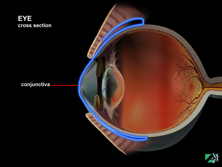
The conjunctiva is a thin, mucus secreting, transparent membrane lining the interior surface of the eyelid and the exposed surface of the sclera, the outer surface of the eyeball. It serves to protect the eye and to lubricate the eyelid
Conjunctival Cul-de-sac
The conjunctiva is the membrane that lines the surface of the eye and the underside of the eyelids. The conjunctival cul-de-sac is the space between these two conjunctivas. It is also known as the conjunctival fold or conjunctival fornix.
Conjunctivitis
Inflammation of the conjunctiva, which is the membrane that lines the eye and eyelid
Conjunctivocystorhinoplasty
Conjunctivocystorhinoplasty, also known as conjunctivocystorhinostomy or CDCR is a surgical procedure for the treatment of epiphora, which is excessive overflow of tears. It involves placement of a stent to bypass the normal lacrimal passages
Conjunctivocystorhinostomy
Same as Conjunctivocystorhinoplasty, surgical procedure to correct excessive production of tears
Continent Ileostomy
Continent ileostomy is a procedure for individuals who have uncontrollable diarrhea and incontinence resulting from previous bowel surgery. The procedure involves creating an internal reservoir and valve made from the small intestine. No external bags are needed. The reservoir is empted by the patient inserting a drainage catheter
Contrecoup Fracture
A skull fracture that occurs distant from the point of impact, for example a blow to the forehead may result in a fracture at the back of the head
Contrecoup Lesion
A type of brain contusion where the contusion to the brain occurs opposite the impact to the skull
Contusion
A contusion is a bruise, an injury involving the cutaneous surface of the skin without any break in the skin
Conus Medullaris
The terminal portion of the spinal cord from which the cauda equina emerges
Coracobrachialis
A muscle extending from the shoulder to the upper arm. It originates from the coracoid process of the scapula at the shoulder and inserts onto the medial surface (inside) of the humerus just above the elbow. It assists flexion and adduction of the arm. The musculocutaneous nerve innervates it
Coracoclavicular Ligament
A shoulder ligament, it helps support the acromioclavicular joint
Coracohumeral Ligament
A ligament in the shoulder, it helps support the shoulder
Coracoid Process
A bony projection from the scapula, which gives attachment to several shoulder ligaments
Coreoplasty
Plastic repair of the iris
Corium
Another name for dermis, one of the layers of the skin, the deeper layer
Cornea
The cornea is the circular transparent part of the front aspect of the eye. It is convex in shape to permit refraction of incoming light waves. It consists of five layers
Corneal Tattooing
Corneal tattooing is a procedure performed to correct cosmetic blemishes on the cornea. A microscopic tattooing instrument may be used to pigment the blemish, or the tattooing may be done manually under a microscope
Coronal Plane
A term used to describe a sectional surface of the body. If you imagine cutting the body in two from head to toe so as to divide into a front and back part, but not necessarily in equal parts, then this is referred to as the coronal plane
Coronal Suture
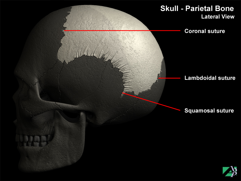
The regions of the skull are divided by tissue and cartilage called sutures. The coronal suture divides the frontal and parietal bones
Coronoid Fossa
A depression in the distal humerus (at the elbow), which the ulna fits into
Coronoid Process of Mandible
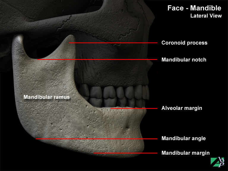
Part of the mandible, it serves as an attachment point for the temporalis muscle
Coronoid Process of Ulna
A bony lip below the olecranon (the tip of the elbow)
Corpus Callosum
The corpus callosum is situated in the middle of the brain. Its serves to connect the two hemispheres of the cerebrum
Corpus Striatum
Masses of grey and white matter within the brain
Corrugator
A facial muscle originating from above the eyebrows and inserting onto the root of the nose. It causes wrinkling between the eyes, (as when a person frowns)
Cortical Bone
The superficial layer of compact bone, which is hard dense bone
Corticosteroid
Corticosteroids are a chemical substance of the steroid group, which have the properties and action of the hormones produced by the adrenal gland. They are used for the treatment of many musculoskeletal disorders because of the anti-inflammatory responses. Corticosteroids may be taken as a topical ointment, orally, by inhalation and by injections
Costal Cartilage
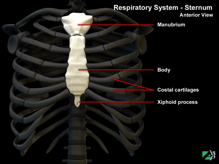
The ribs attach to the sternum via cartilaginous links known as costal cartilages. Apart from being points of attachment, the costal cartilages also play an important role in the elasticity of the chest wall. The first seven pairs of cartilages attach to the sternum, the next three attach to the costal cartilage of the attaché to the cartilage of the seventh rib. The last two attach to the wall of the abdomen
Costochondral
The term relates to both a rib and its costal cartilage
Costoclavicular Ligament
A ligament that supports the sternoclavicular joint
Costovertebral
The term applies to the area where a rib joins a vertebra
Coup Lesion
A type of brain contusion where the contusion to the brain occurs at the site of the impact to the skull
Cranial Fossa
A depression on the inside of the floor of the cranium
Cranial Nerve
A peripheral nerve arising from the brain. There are 12 paired cranial nerves, numbered in Roman numeral style, they are: Olfactory nerve (Cranial Nerve I), Optic nerve (Cranial Nerve II), Oculomotor nerve (Cranial Nerve III), Trochlear nerve (Cranial Nerve IV), Trigeminal nerve (Cranial Nerve V), Abducent nerve (Cranial Nerve VI), Facial nerve (Cranial Nerve VII), Vestibulocochlear nerve (Cranial Nerve VIII), Glossopharyngeal nerve (Cranial Nerve IX), Vagus nerve (Cranial Nerve X), Accessory nerve (Cranial Nerve XI), and Hypoglossal nerve (Cranial Nerve XII)
Cranial Nerve 1 (Olfactory Nerve)
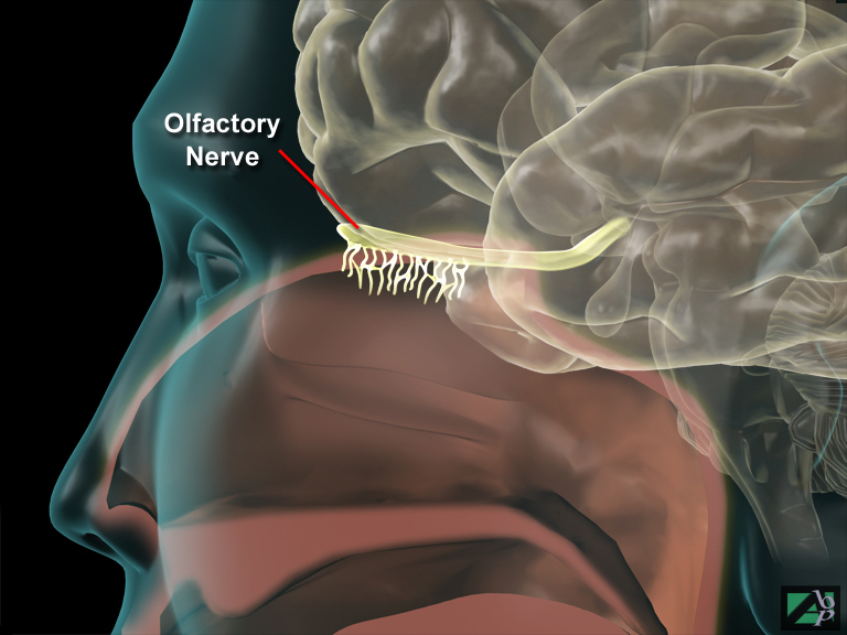
Cranial nerve I is the olfactory nerve. It serves purely sensory functions. Actually, it is not a nerve per-se but rather a sensory fiber tract that relays sensory impulses of smell from the mucous membranes within the nasal cavity. The nerve tract functions as chemoreceptors that respond to chemical particles breathed into the nasal cavity. Sensory reactions are passed along this olfactory tract to the primary olfactory area in the cerebral cortex for dissemination and recognition
Cranial Nerve II (Optic Nerve)
Cranial nerve II is the optic nerve. It is purely a sensory nerve comprising an estimated 1.2 million nerve fibers. It conducts impulses from the rods and cones of the retina in the eye along what is known as the optic chiasma (near the pituitary in the floor of the diencephalon) then through optic tracts to the thalamus then to the visual cortex of the occipital lobes
Cranial Nerve III, (Oculomotor Nerve)
The oculomotor nerve is primarily a motor nerve that innervates muscles of the eyeball and muscles of the iris of the eye to initiate pupil dilation. It also innervates muscles within the ciliary body of the eye to produce lens accommodation (spontaneous adjustment of the eye to focus on near and distant objects)
Cranial Nerve IV, (Trochlear Nerve)
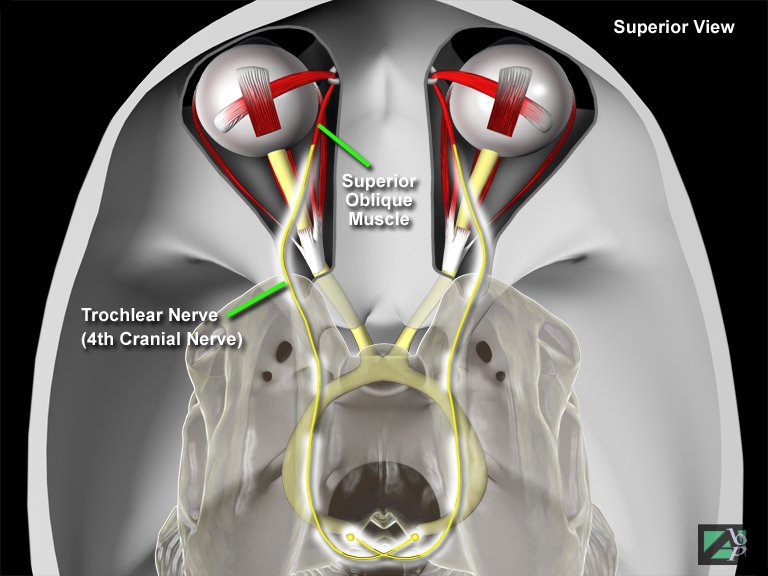
The trochlear nerve is a mixed nerve (motor and sensory functions) that innervates the superior oblique muscle of the eyeball that permits downward rotation of the eyeball
Cranial Nerve V (Trigeminal Nerve)
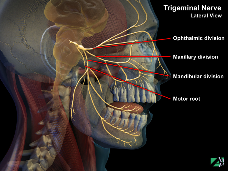
This is a large mixed nerve that has three branches: the ophthalmic branch, maxillary branch and mandibular branch. Although it is a mixed nerve its sensory functions are much more extensive than its motor functions. The motor fibers of the trigeminal nerve are found in the mandibular branch that innervates mouth muscles used in mastication and also other muscles on the floor of the mouth. The sensory fibers of the mandibular branch respond to sensation from the teeth and gums of the lower jaw, parts of the tongue, auricle of the ear and lower part of the face. The ophthalmic sensory branch has sensory fibers that respond to touch, temperature and pain from the anterior half of the scalp, skin of the forehead, upper eyelid, surface of the eyeball, and side of the nose. The maxillary branch responds to sensation from the lower eyelid, palate and portion of the pharynx, teeth and gums of the upper jaw
Cranial Nerve VI (Abducens Nerve)
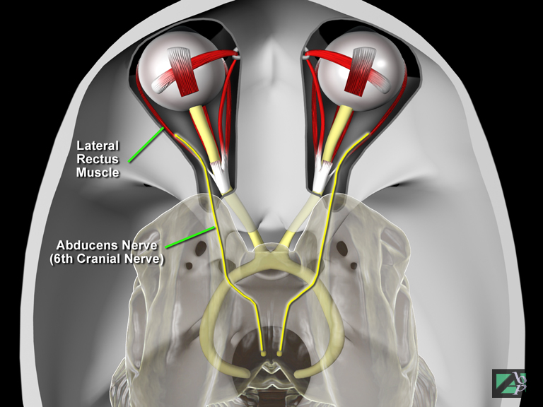
This is a mixed nerve (sensory and motor) that innervates the lateral rectus eye muscle that causes the eyeball to rotate laterally (to the side)
Cranial Nerve VII (Facial Nerve)
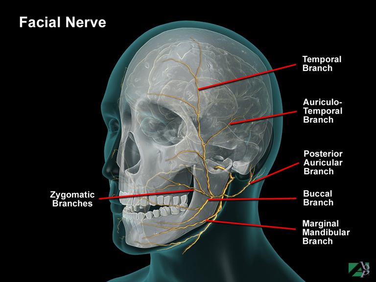
The facial nerve is a mixed nerve. The motor fibers innervate the facial muscles and scalp muscles. Sensory fibers of this nerve supply the sense of taste
Cranial Nerve VIII (Auditory Nerve)
(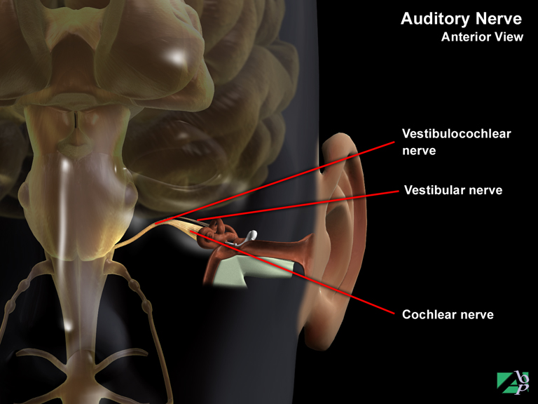
The acoustic nerve, also known as the vestibuloocular nerve is a mixed or composite sensory nerve. It comprises two branches that arise within the inner ear. These are the cochlear division for hearing and the vestibular division for equilibrium and balance
Cranial Nerve IX (Glossopharyngeal Nerve)
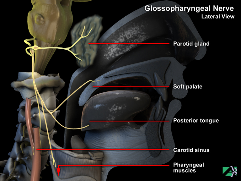
The glossopharyngeal nerve is a mixed sensory and motor nerve that innervates part of the tongue and pharynx. The motor fibers innervate the muscles of the pharynx and the salivary gland to stimulate the swallowing reflex and secretion of saliva. The sensory fibers provide the sensation of taste for bitter and sour taste perception
Cranial Nerve X (Vagus Nerve)
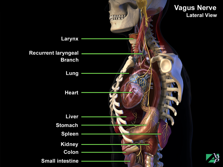
The vagus nerve is also a mixed sensory and motor nerve with an important parasympathetic division to thoracic and abdominal viscera. Through various branches it innervates the muscles of the pharynx, larynx, respiratory tract, lungs, heart, esophagus and some abdominal viscera. One branch of the nerve, the recurrent laryngeal nerve, enables speech
Cranial Nerve XI (Accessory Nerve)
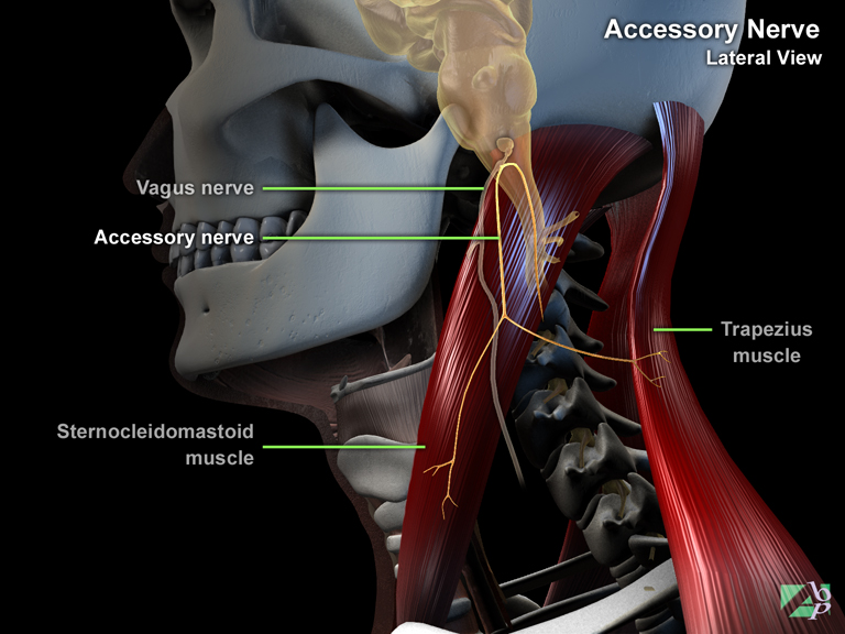
The accessory nerve is primarily a motor nerve though it does have some sensory fibers. This nerve is unique among the cranial nerves in that it has its origins both in the brain and the spinal cord. The cranial motor fibers innervate the skeletal muscles of the palate, pharynx and larynx. The spinal motor fibers of the accessory nerve innervate the muscles that move the head, neck and shoulders
Cranial Nerve XII (Hypoglossal Nerve)
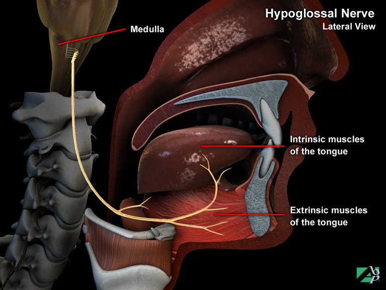
The hypoglossal nerve is a mixed nerve with both motor and sensory fibers. The motor fibers innervate tongue muscles while the sensory fibers monitor the position and function of the muscles
Cranial Puncture
Cranial puncture is a procedure carried out to relieve increased intracranial pressure
Cranial Sutures
These are suture lines between the bones of the skull
Cranial Vault
The upper domed portion of the skull. It comprises the frontal, occipital and parietal bones
Craniectomy
This is a medical procedure in which part of the cranium (skull) is removed
Cranioplasty
Cranioplasty is a neurological procedure to repair a bony defect, or deformity in the skull
Craniosacral Technique
A type of osteopathic manipulation
Craniotomy
This is a medical procedure in which a surgical incision is made into the skull
Cranium
Another term for skull
Crepitus
Describes a crackling sound on joint movement
Crest
A ridge
Cribriform Plate
Part of the ethmoid bone which forms the floor of the skull, the cribriform plate is the horizontal section of the bone
Cricoid Cartilage
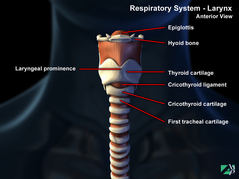
A round cartilage situated between the upper end of the pharynx and the lower end of the larynx
Cricopharyngeal Myotomy
Cricopharyngeal myotomy is a surgical procedure performed for the treatment of swallowing difficulties caused by a dysfunctional esophageal sphincter muscle. The sphincter muscle is cut via an external incision in the neck
Cross Union
Cross union or as it is more properly known, radioulnar synostosis, is a rare abnormal bony union between the proximal radius and ulna at the elbow. This cross union prevents rotation of the forearm
Crown
The part of a tooth that projects above the gum line
Cryotherapy
Cryotherapy is a surgical technique that utilizes freezing tissue to remove unwanted tissue. It has widespread use in ophthalmic surgery and is also used to destroy warts and skin lesions and other body lesions
CSF Otorrhea
Leakage of cerebrospinal fluid from the ear
CSF Rhinorrhea
Leakage of cerebrospinal fluid from the nose
Cubital Tunnel Syndrome
Cubital tunnel syndrome is an entrapment syndrome of the ulnar nerve at the elbow. It may be due to trauma involving fractures or dislocations of the elbow or from overuse injury or may be occupationally related
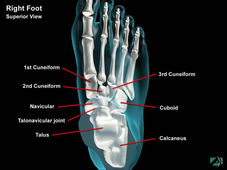
Cuboid Bone
The cuboid, which is six sided is located on the lateral or outer side of the foot. The posterior surface connects with the calcaneus, while it's anterior or front surface connects to the 4th and 5th metatarsals (long bones of the foot). Its medial surface (towards the inner side of the foot or big toe side) connects to the navicular and one of the cuneiform bones
Cuneiform
One of three bones of the foot. They lie between the navicular (behind) and the first three metatarsal bones in front
Cusp
Refers to either part of a valve of the heart or the rounded projection on the chewing surface of a tooth
Cuspid
A tooth with one point, or cusp, there are four cuspids lying on either side of the incisor teeth
Cutaneous
Pertaining to the skin
Cyanosis
A bluish tinge to the skin. It usually signifies a lack of oxygen in the blood
Cyst
A fluid filled sac
Cystitis
Inflammation of the urinary bladder
Cystoscopy
An examination of the interior of the bladder using a cystoscope, which is a tube with an optical lens
Cystostomy
A cystostomy, which can be temporary or permanent, is a procedure whereby a catheter is placed through the abdominal wall into the bladder as a form of urinary diversion. It is performed because of blockage or stricture of the urethra, the canal that carries urine from the urinary bladder to the outside of the body