Medical Glossary - Letter P
This medical glossary of terms beginning with the letter "P" contains the more common medical terms one might expect to encounter in a medical report or in hospital notes. The glossary is intended as a quick reference only; many of the terms are also referenced and illustrated in more detail in the medical libraries, to which you should refer for more detailed information.
Palate
The roof of the mouth. It is divided into a soft and hard palate. The roof at the back of the mouth is the soft palate; the rest is the hard palate
Palatine Bone
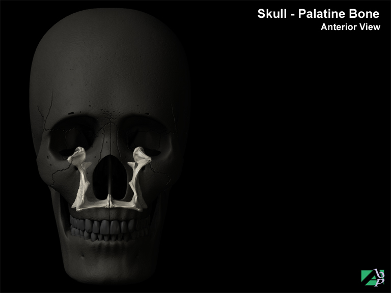
The palatine bones are bones of the face; there are two, a left and right palatine bone. They form part of the roof of the back of the mouth, the floor of the orbits (eye sockets) and the floor and sidewalls of the nasal cavities
Palmar
Referring to the palm of the hand
Palmar Artery
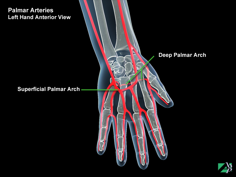
This is a continuation of the radial artery that joins the ulna artery in the palm of the hand and which then forms the network of digital arteries that extend into the fingers
Palmaris Brevis
A muscle in the superficial aspect of the palm of the hand, which assists the hand in gripping objects, it is innervated by the ulnar nerve
Palmaris Longus
A muscle of the arm that originates from the medical epicondyle of the humerus at the elbow and travels down the forearm and inserts onto the palm of the hand. It assists wrist flexion. The median nerve innervates it
Palsy
A synonym for paralysis
Pancreas
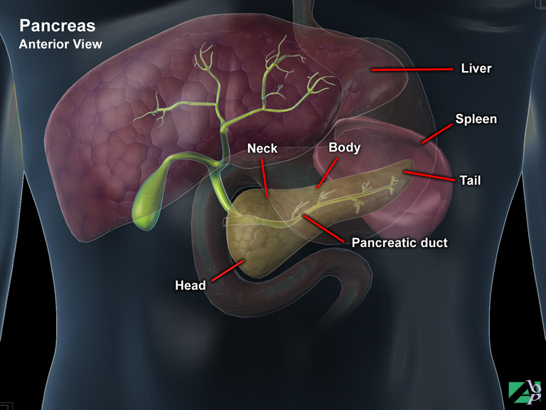
The pancreas is located below the stomach between the liver and the spleen and behind a portion of the large intestine, roughly at the level of the twelfth thoracic vertebra. It is approximately 12.5 centimeters (5 inches) long and 2.5 centimeters (1 inch) thick. It is comprised of four sections: a head, neck, body and tail. The pancreas is part of the endocrine system i.e., part of the glandular system. Its function is vital to the metabolic processes of the body. Its secretions are essential to intestinal digestion and to insulin production that enables the body to absorb and metabolize (transform) glucose. The pancreas has two separate functional systems known as the pancreatic exocrine system and the pancreatic endocrine system. The pancreatic exocrine system produces three enzymes that pass through a duct system and which are secreted into the duodenum, this is referred to as pancreatic juice. This juice contains water, bicarbonate and a variety of digestive enzymes that are used by the duodenum to digest food passed through by the stomach. About 98% of the pancreas's cell tissues are engaged in production of these digestive enzymes. The pancreatic endocrine system functions to produce insulin and other hormones. The insulin producing cells are located in an area known as the islets of Langerhans. Insulin regulates the body's uptake of glucose
Pancreatitis
Inflammation of the pancreas. It may be acute or chronic
Pancreatogram
Pancreatogram is an x-ray of the biliary system (pancreas and bile ducts) and is usually performed for the detection of gallstones, or narrowing due to other conditions. An endoscope (fiberoptic tube) is passed through the mouth, esophagus and stomach and a smaller tube is placed into the opening of the common bile duct (from the gallbladder). X-ray dye is injected into the ducts to demonstrate any abnormalities
Paresthesia
An abnormal sensation involving a sensation of tingling, burning and sometimes tickling
Paralytic Ileus
Paralytic ileus is paralysis of the bowel. It may result after abdominal surgery, spinal fractures and retroperitoneal hemorrhage or from abdominal trauma
Paraplegia
Paralysis of both lower limbs and partial or total loss of urinary and bowel function due to spinal cord disease or injury
Parietal Bone
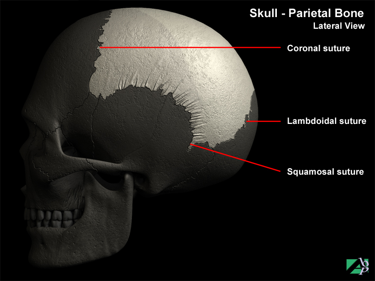
A cranial bone, there are in fact two, one on each side of the skull that form the vault and part of the side walls of the skull
Parietal Lobe
The parietal lobe lies behind the frontal lobe. The cortex (grey matter) of the parietal lobes is concerned with the primary and secondary sensory functions and complex association mechanisms involved in higher-level integration and, in the dominant hemisphere, speech
Parotid Gland
A salivary gland, there are two situated on either side of the head just below the ear
Parumbilical
Parumbilical means near or around the navel
Pars Interarticularis
The pars interarticularis or simply pars, is a narrow piece of bone that lies between the superior and inferior articulating processes of the vertebra. The defect may be found at any level but most commonly occurs in the lumbar spine, particularly at L5
Patella
The patella, or as it is more commonly known, the kneecap, is a rounded triangular shaped bone, which serves to protect the knee joint and to strengthen the quadriceps tendon. It is a sesamoid type of bone, which means that it is a bone that is situated within a tendon, in this case the quadriceps tendon. At birth, the patella is actually formed entirely of cartilage, which later ossifies (converts) into bone. The patella connects with the femoral condyles and glides up and down a trough or track on the femur; it constantly changes position with knee movements.
Patella Retinaculum
Fibrous tissue around the patella
Patella Stabilization
Patella stabilization is an operation to secure the patella in cases of recurrent dislocation. The procedure involves transfer of the patella ligament. The patella ligament with the segment of bone into which it is inserted is freed and reattached further medially and distally. An alternative to this is the lateral half of the ligament is detached, threaded through the medial half and reattached more medially and distally. Both procedures prevent further dislocation of the patella
Patellectomy
Removal of the patella
Patellofemoral
Relating to both the femur and the patella
Pathologic
Diseased, abnormal
Pectineus
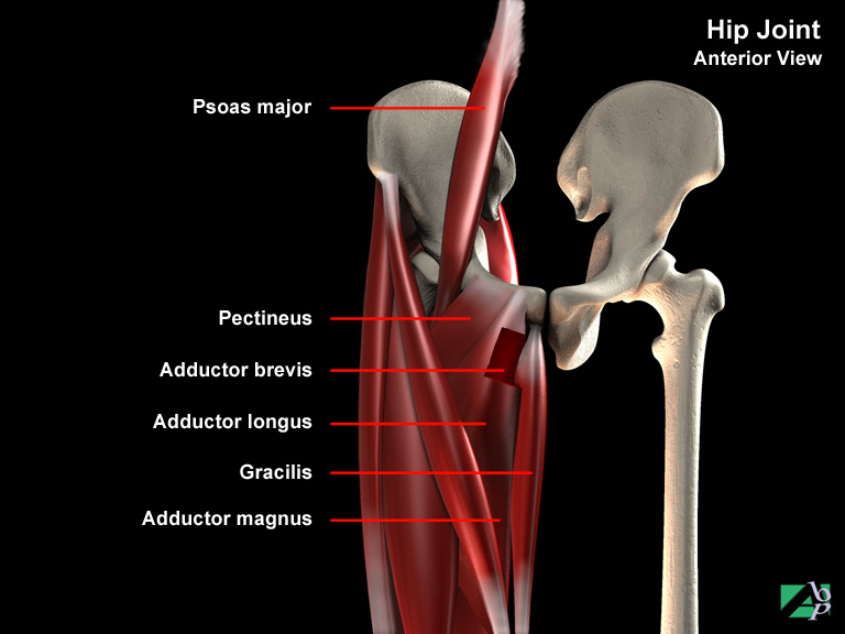
A muscle of the hip that originates from the pelvis and inserts onto the femur just below the lesser trochanter. It assists flexion, adduction and medial rotation of the hip. The femoral nerve innervates it
Pectoral
Relating to the chest
Pectoral Girdle
The shoulder girdle
Pectoralis Major
A major muscle of the chest wall that originates from the clavicle and sternum and attaches onto the humerus and deltoid tuberosity. It flexes and adducts the arm and assists with medial rotation of the arm
Pectoralis Minor
A muscle of the chest wall that originates from the 3rd, 4th and 5th ribs and inserts onto the coracoid process of the scapula. It assists elevation of the ribs and protraction of the scapula
Pelvic
Relating to the pelvis
Pelvis
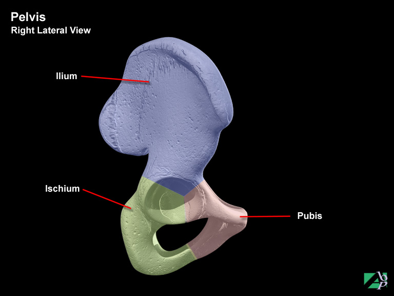
The pelvis is formed by two hipbones called ossa coxa, which are united at the rear by the symphysis pubis. It is attached posteriorly to the vertebral sacrum and coccyx. The pelvis, or pelvic girdle supports the weight of the body and provides protection for the lower abdominal viscera and the reproductive organs. Each ossa coxa comprises three separate bones, which fuse together in adulthood. These bones are the ilium, ischium and pubis. On the side of the ossa coxa where the three bones fuse is a large circular depression, the acetabulum, which articulates with the head of the femur to form the hip joint. The ilium is the largest of three bones and is at the top of the ossa coxa. It has a crest and four spines for muscle attachments. The iliac crest is the prominent or protruding part of the hipbone. The spines are named by their position i.e., anterior superior iliac spine, anterior inferior iliac spine, posterior superior iliac spine and posterior inferior iliac spine. Below these spines are roughened irregular surfaces that serve as attachments for the sacroiliac ligament and gluteal muscles. These are the iliac fossa, iliac tuberosity and gluteal lines. Behind and below the ilium is the ischium. This is comprised of a spine, a sciatic notch, a tuberosity (a bony eminence), a ramus (an extension of bone) and an obturator foramen (opening). The pubis is the lower portion of the ossa coxa and forms the joint between the two ossa coxa known as the symphysis pubis. It comprises a body, and two rami known as the superior ramus and inferior ramus
Percutaneous
Through the skin
Periosteum
A membrane that lines the surface of a bone
Periostitis
Periostitis is inflammation of the periosteum, which is a membrane that surrounds the surfaces of bones. Trauma can be the cause of the inflammation as can excessive exercise as in running. The tibia is the most common site for periostitis. Periostitis can also result from an earlier bout of osteomyelitis
Peritoneal Adhesions
Peritoneal adhesions, or as they may be called, stomach adhesions, are scar tissue that has formed inside the abdomen. They can form in response to traumatic injury, surgery or inflammatory diseases inside the abdomen. They can form on the outside of the abdominal wall, the peritoneum, on or between loops of the bowel or between any of the abdominal organs. They are frequently the cause of bowel obstruction
Peritoneal Cavity
Same as abdominal cavity, the space in which the abdominal organs are contained
Peritoneum
The peritoneum is a thin membrane that lines the abdominal cavity and covers the organs within the abdominal cavity. It is lubricated by serous fluid (thin fluid) that permits the abdominal organs to move against each other and the abdominal wall. The peritoneum that lines the abdominal cavity is called the parietal peritoneum. That which lines the organs is known as the visceral peritoneum
Peritonitis
Peritonitis is a life threatening condition, which requires urgent medical attention. It is an inflammation of the peritoneum, the thin membrane that lines the abdominal cavity and the organs that lie within. It is most commonly caused by the bacteria E Coli, Klebsiella and Proteus. It usually results from an external source such as an injury or inflammation from another abdominal organ. It may also follow surgical procedures. Common causes are appendicitis, bowel perforations, perforated ulcers, trauma and infections
Perivesical
Refers to around the bladder
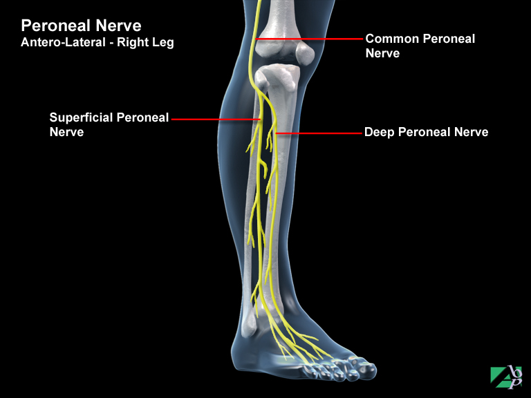
Peroneal Nerve
The peroneal nerve, or as it sometimes is referred to the common peroneal nerve, is a continuation of the sciatic nerve. At the knee, it separates into two branches, the superficial and deep peroneal nerve. It is a mixed nerve (motor and sensory). It provides motor impulses to the muscles of the calf of the leg and sensation to the dorsum of the foot and the front of the leg. It is particularly vulnerable to injury with associated knee injuries
Peroneus Brevis
A muscle on the outer side of the leg that originates from the shaft of the fibula and inserts onto the base of the 5th metatarsal of the foot. It assists dorsiflexion and eversion of the foot. The peroneal nerve innervates it
Peroneus Longus
A long muscle on the outer side of the leg, originating from the upper ends of the tibia and fibula and inserting onto the cuneiform and 1st metatarsal bones of the foot. It assists eversion of the foot and extension of the ankle joint. The peroneal nerve innervates it
Peroneus Tertius
A muscle on the outside of the lower leg that originates from the fibula and inserts onto the base of the 5th metatarsal. It assists extension and eversion of the foot. The peroneal nerve innervates it
Pertrochanteric Fracture
A fracture of the femur in which the fracture line extends through the trochanter region of the femur
Pes anserinus
A tendon that anchors the sartorius, gracilis and semitendinosus muscles onto the tibia
Pes Anserinus Tendinitis
This term describes tendinitis (inflammation of a tendon) of the pes anserinus tendon, which attaches to the tibial tuberosity of the knee. It is an expanded tendon formed by three muscles, the sartorius, gracilis and semitendinosus muscles
Pes cavus
A foot deformity in which the arch of the foot is curved greater than normal resulting in the heel being abnormally elevated
Petit mal
A milder form of epileptic seizure with short loss of consciousness but no epileptic convulsions
Phalange
A digit, a finger or toe, refers to the entire digit
Phalangectomy
Phalangectomy is the surgical removal of part of a phalanx
Phalanx
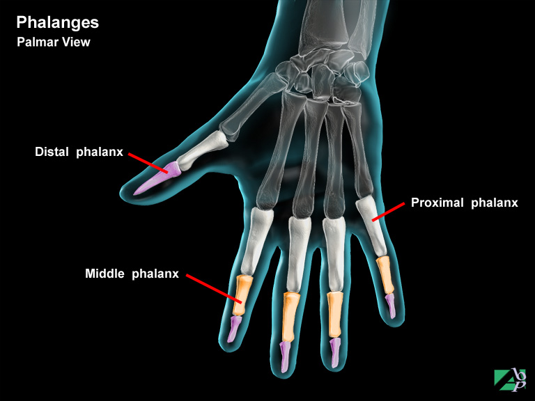
Part of a phalange. With the exception of the thumb and big toe the fingers and toes are comprised of three phalanxes. The tip or end phalanx is the distal or terminal phalanx; the nearest phalanx (the part closest to the foot or hand) is the proximal phalanx. The section in between is the middle phalanx. The big toe and thumb have only two phalanges; they do not have a middle phalanx
Phantom Limb Pain
Phantom limb pain is the term used to describe the feeling an amputated limb is still attached. It is caused by irritation of the stump of the nerve that innervated the amputated part that was severed in the amputation. Phantom sensations include pain, touch, pressure, wetness, itching or fatigue. The sensation is strongest in the immediate post-amputation period and then recedes or disappears completely, but in some cases may persist indefinitely. It is a common post amputation complication
Pharyngoscopy
This is an instrument examination of the pharynx using a pharyngoscope, which is a specially designed instrument that allows visualization of the interior of the pharynx
Pharyngeal
Refers to the pharynx or throat area
Pharyngeal Diverticulectomy
Pharyngeal diverticulectomy is a surgical procedure performed for the treatment of a pharyngeal diverticulum. A diverticulum is a herniation (protrusion) of the mucous lining of the pharynx that protrudes through the pharynx. Diverticulectomy is the surgical removal of a diverticulum
Pharyngotomy
Pharyngotomy is a surgical incision into the pharynx. It may be made through several approaches including from within, (internal pharyngotomy), externally or laterally (from the side)
Pharynx
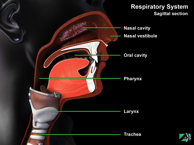
The pharynx plays a dual role; it is part of the respiratory system and also functions as part of the digestive system. It is a funnel shaped passageway that connects the back opening of the nose to the esophagus, which transports food to the stomach and in respiration, tunnels air to the larynx and trachea. The pharynx is divided into three regions, the nasopharynx, oropharynx and the laryngopharynx. The nasopharynx is the uppermost division that extends from the back of the nose to the roof of the mouth. This is a respiratory division. The nasopharynx also has connections with the middle ear cavities via the paired auditory and Eustachian tubes. The oropharynx or throat is the part that extends from the roof of the mouth to the level of the hyoid bone (just above the larynx). The base of the tongue forms the front of the oropharynx. Along the rear wall are the paired palatine tonsils. This portion of the pharynx performs both respiratory and digestive functions. The laryngopharynx portion extends from the hyoid bone to the esophagus at which point the respiratory and digestive systems become distinct. Food is passed to the esophagus and air to the trachea
Phlebitis
Inflammation of a vein
Phlebography
Phlebography is an x-ray examination of veins. The x-ray picture known as a phlebogram is taken after the injection of a radiopaque material into the vein
Phlebotomy
The surgical cutting into a vein
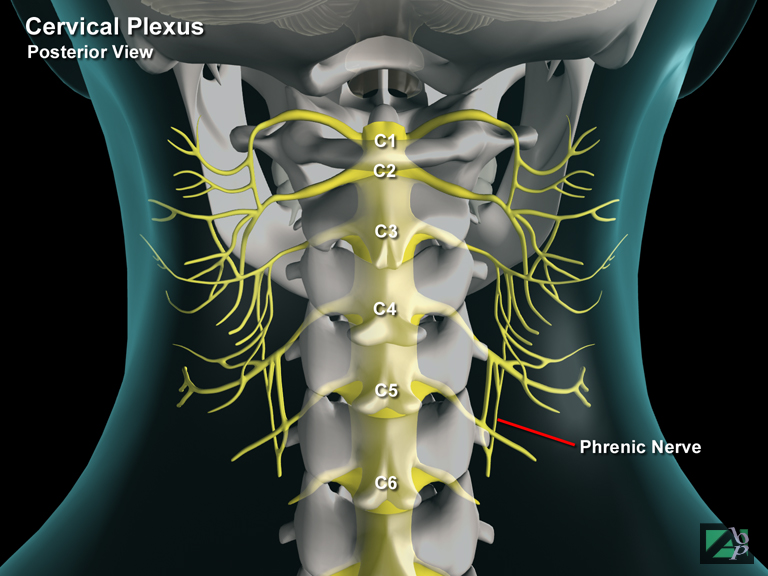
Phrenic Nerve
The phrenic nerves (it is a paired nerve) innervates the diaphragm and thus assists in the act of inhaling and expelling air from the lungs
Pia Mater
The innermost layer of the meninges that cover the brain and the spinal cord, the other layers are the arachnoid and the dura mater, the arachnoid is the middle layer and the dura is the outer layer
Pinna
The fleshy external part of the ear
Piriformis
A muscle that originates in the lower back from the sacrum and inserts onto the femur at the greater trochanter. It assists lateral rotation of the hip and also to stabilize the hip
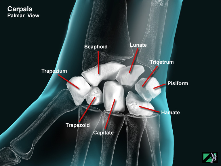
Pisiform
A bone of the wrist, it is pea shaped and connects with the triquetrum and has strong tendon attachments.
Pituitary Gland
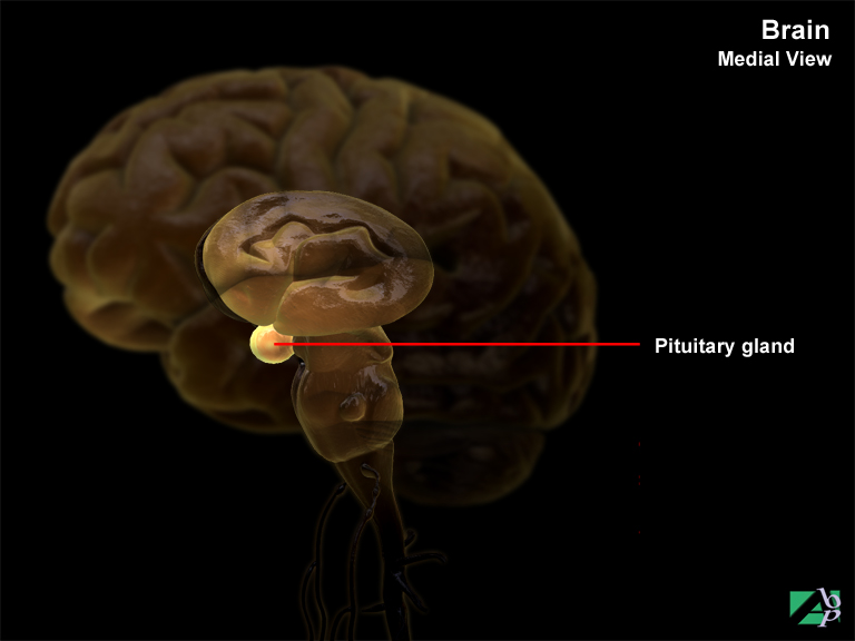
The pituitary gland is a small structure about the size of a pea on the underside of the brain. Its main function is the production of a number of hormones among these being growth hormones that stimulates growth of the body and which also influence the metabolism of carbohydrates, fats and proteins. It also produces hormones that control the function of the thyroid and adrenal glands and hormones that stimulate the gonads, ovaries and testicles. In addition it stimulates the production of milk. Other hormones produced affect the contraction of the smaller blood vessels and the output of urine
Plantar
Refers to the sole or underside of the foot
Plantar Fasciitis
Inflammation of the fascia of the heel pad
Plantaris
A muscle of the leg originating from the supracondylar region of the femur just above the knee and travelling down the inside of the leg and inserting onto the heel. It assists knee flexion and plantar flexion of the foot. The tibial nerve innervates it
Pleural Effusion
Fluid within the chest, or more specifically in the pleural cavity, between the pleural lining of the lung and the pleural lining of the chest wall
Pleuroperitoneal Shunt
Pleuroperitoneal refers to the pleural cavity and the peritoneal cavity, which are the two cavities of the body separated by the diaphragm. A shunt is a surgically implanted artificial channel created for the purpose of draining accumulated fluid. Pleuroperitoneal shunts are a form of treatment for the relief of accumulated pleural fluid, usually caused by pleural effusion. The shunts have a subcutaneous pumping chamber that is connected to the pleural space at one end and to the peritoneal cavity at the other. This allows fluid to be pumped directly from the pleural space to the peritoneum
Plexus
A bundle of nerves that originate from the spinal cord, the major plexus of the body are the cervical plexus, brachial plexus, lumbar plexus and sacral plexus
Pneumoencephalogram
A pneumoencephalogram is an x-ray of the brain taken after the injection of air into the ventricles of the brain and other areas occupied by cerebrospinal fluid. Some risks are attached to the procedure and today it is regarded as less reliable than CT and MRI for scanning potential abnormalities of the brain
Pneumohemothorax
The presence of air and blood on the pleural cavity, the space between the pleura lining the lungs and the pleura lining the chest wall
Pneumonia
Pneumonia is inflammation and congestion of the lung or lungs. It may follow chest trauma or be a complication of general infirmity while an individual is recovering from serious injury. The elderly are particularly prone to developing pneumonia while debilitated with another illness
Pneumothorax
Air in the pleural space or cavity. Air in the space between the pleural lining of the lung and the pleural lining of the chest wall
Popliteal Cyst
A cyst filled with synovial fluid, also known as a Baker's Cyst. It can be caused by a number of conditions e.g., irritation of a bursa, cartilage tears or by osteoarthritis within the joint. Symptoms include swelling behind the knee, pain and difficulty straightening the leg
Polydipsia
Excessive thirst
Polyuria
Excessive urination
Pons
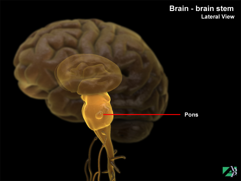
The pons is part of the brain stem derived from the Latin "pons" meaning bridge. It is the crossover point for ascending sensory and descending motor tracts linking higher and lower brain centers, and for transverse fiber pathways entering and leaving the cerebellum on each side. Its significance lies in the fact three cranial nerves arise from it and along with the medulla oblongata it regulates the rate and depth of breathing. The cranial nerves are the sixth cranial nerve, the abducens nerve, seventh cranial nerve, the facial nerve and the eighth cranial nerve, the vestibuloocular nerve
Popliteal
Refers to the back of the leg, particularly the region in the back of the knee
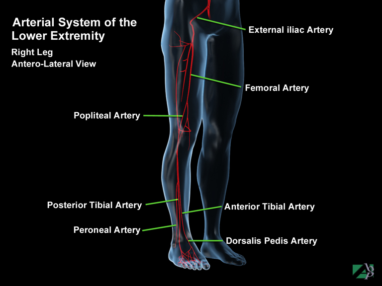
Popliteal Artery
The popliteal artery is a continuation of the femoral artery and commences at the knee; it supplies blood to the knee joint. As it descends below the knee, it divides into the anterior and posterior tibial arteries
Popliteus
A muscle of the knee originating from the tibia and inserting onto the lateral femoral condyle and the lateral meniscus in the knee joint. It assists lateral rotation of the femur on the tibia and retracts the lateral meniscus
Post Concussive Syndrome
Post Concussive syndrome is a term used to define a bunch of ill-defined symptoms that follow concussive head injuries. It is a contentious diagnosis that many clinicians question. It may also be referred to as posttraumatic brain syndrome, mild traumatic brain injury, posttraumatic syndrome and posttraumatic headache syndrome. Many symptoms have been attributed to the syndrome but the primary symptoms are headache, vertigo and difficulty in concentration, depression, apathy and anxiety. The cause of the syndrome is also in dispute, whether it is psychological in nature or whether there is organic basis for the symptoms. What is clear is that there are few organic signs of the syndrome
Posterior
Behind
Posterior Ischiofemoral Ligament
A ligament that supports the hip joint
Posterior Spinal Fusion
A type of vertebral fusion used to stabilize the spine following an injury that has left the affected area unstable. In posterior fusion the spinous and articular processes of the vertebra are removed and strips of bone graft are laid along the lamina and the area is then packed with bone chips
Posterior Sternoclavicular Ligament
A ligament that supports the sternoclavicular joint
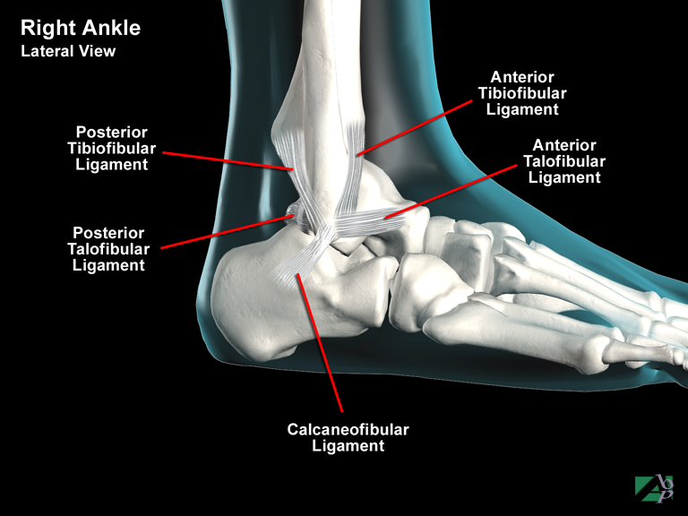
Posterior Talofibular Ligament
A ligament in the ankle that helps support the ankle joint
Posterior Tibial Artery
The posterior tibial artery descends from below the knee down the rear of the leg and provides blood to the muscles of the lower leg and foot. The posterior tibial artery divides at the ankle to become the lateral and medial plantar arteries
Posttraumatic Epilepsy
Although there is a nexus between trauma and epilepsy, the exact reason for this isn't clear. The risk however is known to depend on certain factors. There is a correlation between the severity of head injury and the risk of developing epilepsy. The type of head injury has also been recognized to be a factor, and the risk of epilepsy in the future also depends on the duration of seizure free time following the accident. It is also believed adults are at greater risk of developing posttraumatic epilepsy than children. Posttraumatic epilepsy is generally discussed as being immediate, early or late based
Immediate type occurs straight after trauma or within an hour of trauma. Most often the convulsion is of the clonic type. However the incidence of immediate posttraumatic epilepsy is very low. It is also believed that individuals who do suffer immediate posttraumatic epilepsy are not at increased risk of suffering further episodes
Early posttraumatic epilepsy describes convulsions (usually without loss of consciousness) within the first week of trauma and is more commonly seen in children than adults. Additional risk factors for early onset are if the patient suffered an intracranial hematoma or a depressed skull fracture. Early onset greatly increases the risk of a subsequent chronic disorder
Late onset describes a convulsion if it occurs after the first week. Many of those that suffer late onset suffer partial complex type epilepsy. Most people that suffer late onset do so within 12 months of the trauma but it may be delayed years
Four risk factors have been identified with late posttraumatic epilepsy. They are posttraumatic amnesia that lasts more than 24 hours, depressed skull fracture, intracranial hematoma and if the head injury involved penetrating trauma
Posttraumatic Stress
Posttraumatic stress disorder (PTSD) is a disorder that develops in some people following exposure to a traumatic event. Not every traumatized person develops chronic or even acute PTSD. PTSD can be triggered by direct or indirect exposure to death, serious injury or violence. The threat of death, injury or violence may also be sufficient to trigger PTSD. For a diagnosis of PTSD, symptoms persist at least one month. The symptoms must cause functional impairment or distress and cannot be the result of substance abuse, medication or illness.
A diagnosis of PTSD is associated with at least one re-experiencing symptom such as flashbacks, nightmares, racing heart, sweating or bad thoughts. PTSD criteria also require the presence of an avoidance behavior which may include avoiding a location, activity or thought. At least two arousal and reactivity symptoms are present, including being easily startled, feeling tense or “on edge,” having difficulty sleeping and having angry outbursts. The criteria also require at least two cognition and mood symptoms, such as trouble remembering key features of the traumatic event, negative thoughts about oneself or the world, distorted feelings like guilt or blame or loss of interest in enjoyable activities.
The National Institute of Mental Health (NIMH) is the lead federal agency for research on mental disorders. NIMH is one of the 27 Institutes and Centers that make up the National Institutes of Health (NIH), the largest biomedical research agency in the world. NIH is part of the U.S. Department of Health and Human Services (HHS). The information below regarding Signs and Symptoms and Treatments and Therapies is from NUMH.
Signs and Symptoms
Not every traumatized person develops ongoing (chronic) or even short-term (acute) PTSD. Not everyone with PTSD has been through a dangerous event. Some experiences, like the sudden, unexpected death of a loved one, can also cause PTSD. Symptoms usually begin early, within 3 months of the traumatic incident, but sometimes they begin years afterward. Symptoms must last more than a month and be severe enough to interfere with relationships or work to be considered PTSD. The course of the illness varies. Some people recover within 6 months, while others have symptoms that last much longer. In some people, the condition becomes chronic.
A doctor who has experience helping people with mental illnesses, such as a psychiatrist or psychologist, can diagnose PTSD.
To be diagnosed with PTSD, an adult must have all of the following for at least 1 month:
· At least one re-experiencing symptom
· At least one avoidance symptom
· At least two arousal and reactivity symptoms
· At least two cognition and mood symptoms
Re-experiencing symptoms include:
· Flashbacks—reliving the trauma over and over, including physical symptoms like a racing heart or sweating
· Bad dreams
· Frightening thoughts
Re-experiencing symptoms may cause problems in a person’s everyday routine. The symptoms can start from the person’s own thoughts and feelings. Words, objects, or situations that are reminders of the event can also trigger re-experiencing symptoms.
Avoidance symptoms include:
· Staying away from places, events, or objects that are reminders of the traumatic experience
· Avoiding thoughts or feelings related to the traumatic event
Things that remind a person of the traumatic event can trigger avoidance symptoms. These symptoms may cause a person to change his or her personal routine. For example, after a bad car accident, a person who usually drives may avoid driving or riding in a car.
Arousal and reactivity symptoms include:
· Being easily startled
· Feeling tense or “on edge”
· Having difficulty sleeping
· Having angry outbursts
Arousal symptoms are usually constant, instead of being triggered by things that remind one of the traumatic events. These symptoms can make the person feel stressed and angry. They may make it hard to do daily tasks, such as sleeping, eating, or concentrating.
Cognition and mood symptoms include:
· Trouble remembering key features of the traumatic event
· Negative thoughts about oneself or the world
· Distorted feelings like guilt or blame
· Loss of interest in enjoyable activities
Cognition and mood symptoms can begin or worsen after the traumatic event, but are not due to injury or substance use. These symptoms can make the person feel alienated or detached from friends or family members.
It is natural to have some of these symptoms after a dangerous event. Sometimes people have very serious symptoms that go away after a few weeks. This is called acute stress disorder, or ASD. When the symptoms last more than a month, seriously affect one’s ability to function, and are not due to substance use, medical illness, or anything except the event itself, they might be PTSD. Some people with PTSD don’t show any symptoms for weeks or months. PTSD is often accompanied by depression, substance abuse, or one or more of the other anxiety disorders.
Treatments and Therapies
The main treatments for people with PTSD are medications, psychotherapy (“talk” therapy), or both. Everyone is different, and PTSD affects people differently so a treatment that works for one person may not work for another. It is important for anyone with PTSD to be treated by a mental health provider who is experienced with PTSD. Some people with PTSD need to try different treatments to find what works for their symptoms.
Medications
The most studied medications for treating PTSD include antidepressants, which may help control PTSD symptoms such as sadness, worry, anger, and feeling numb inside. Antidepressants and other medications may be prescribed along with psychotherapy. Other medications may be helpful for specific PTSD symptoms.
Psychotherapy
Psychotherapy (sometimes called “talk therapy”) involves talking with a mental health professional to treat a mental illness. Psychotherapy can occur one-on-one or in a group. Talk therapy treatment for PTSD usually lasts 6 to 12 weeks, but it can last longer. Research shows that support from family and friends can be an important part of recovery.
Many types of psychotherapy can help people with PTSD. Some types target the symptoms of PTSD directly. Other therapies focus on social, family, or job-related problems. The doctor or therapist may combine different therapies depending on each person’s needs.
Effective psychotherapies tend to emphasize a few key components, including education about symptoms, teaching skills to help identify the triggers of symptoms, and skills to manage the symptoms. One helpful form of therapy is called cognitive behavioral therapy, or CBT. CBT can include:
· Exposure therapy. This helps people face and control their fear. It gradually exposes them to the trauma they experienced in a safe way. It uses imagining, writing, or visiting the place where the event happened. The therapist uses these tools to help people with PTSD cope with their feelings.
· Cognitive restructuring. This helps people make sense of the bad memories. Sometimes people remember the event differently than how it happened. They may feel guilt or shame about something that is not their fault. The therapist helps people with PTSD look at what happened in a realistic way.
There are other types of treatment that can help as well. People with PTSD should talk about all treatment options with a therapist. Treatment should equip individuals with the skills to manage their symptoms and help them participate in activities that they enjoyed before developing PTSD.
Pressure Dressing
A dressing applied to exert pressure on a wound
Pressure Point
An area on the skin where nerve endings are contained
Pressure Sore
Another term for decubitus ulcer, an ulceration caused by prolonged pressure on the skin, usually the buttock or heel areas. Bed ridden individuals are especially prone to developing pressure sores
Pronator Quadratus
A muscle in the lower forearm originating from the distal shaft of the ulna and inserting onto the shaft of the radius and the interosseus membrane, a membrane that binds the two bones. It assists pronation of the forearm and to keep the radius and ulna in opposition to each other
Pronator Teres
A muscle of the arm originating from the humerus just above the elbow and inserting onto the radius about mid-forearm. It assists pronation of the forearm. The median nerve innervates it
Proximal
A term often used to describe part of the body or more particularly part of a limb. Proximal means nearest, i.e., nearest the body. For example long bones are often referred to as having a proximal end and a distal end. In the case of the femur the proximal end is the end nearest the hip or lower trunk, whereas the distal end is the end near the knee. In another example the proximal humerus is the part of the humerus nearest the shoulder; the distal end is the end near the elbow
Proximal Interphalangeal Joint
A joint of a toe or finger, in the case of the fingers it is the joint between the metacarpal (hand) bone and the finger; in the toes it is the joint between the metatarsal (foot) bone and the toe. The term is commonly abbreviated to PIP joint
Pruritus
Intense itching
Pseudarthrosis
This term describes a fracture, which has united resulting in abnormal mobility at the fracture site; it is sometimes known as a false joint
Pseudophakos
A Pseudophakos is an artificial implanted intraocular lens (IOC)
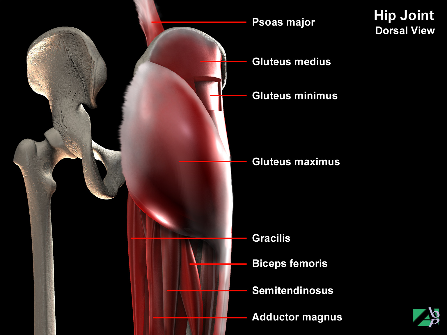
Psoas Major
A hip muscle. It originates from the lumbar region and attaches to the lesser trochanter of the femur. It assists flexion at the hip joint and external rotation of the thigh
Psoas Minor
A pelvic muscle. It originates from the 12th thoracic vertebra and inserts onto the pelvis. It assists flexion of the lower spine and to tilt the pelvis
Psychogenic Overlay
The term describes an exaggeration of symptoms due to emotional factors
Psychogenic Pain
Pain that has no physical or organic basis
Pterygium
A pterygium is a wedged shaped fibrovascular growth of conjunctiva that extends onto the cornea. It is thought that it might develop because of prolonged exposure to ultra violet rays
Ptosis
Drooping of the upper eyelid
Pubofemoral Ligament
A ligament that supports the hip joint
Pulmonary
Relating to the lungs
Pulmonary Artery
The pulmonary artery commences at the right ventricle of the heart and then divides into a left and right branch, which carries blood to the lungs to oxygenate and purify
Pulmonary Contusion
Pulmonary contusion describes bruising of the lung tissue. The vast majority of cases occur in motor vehicle accidents. It occurs usually from blunt trauma and severe decelerating forces. Contusion results in bleeding within the lung tissue causing edema, which affects the gas exchange that takes place within the affected lung. If the contusion is extensive this results in blood from the injured lung being returned to the heart without being oxygenated and with carbon dioxide still present. Respiratory failure can result
Pulmonary Embolism
Pulmonary embolism is a complication of venous thrombosis. In the majority of cases the emboli originates in the deep veins of the lower extremity. Most pulmonary emboli come from blood clots that form in the proximal deep veins, which are the veins above the calf muscles. Trauma patients with the highest risk of pulmonary embolism include those with severe head injuries and coma, spinal cord injuries and those with fractures of the pelvic and long bones. Surgical procedures can also lead to pulmonary embolism
Pulmonary Edema
Pulmonary edema is a life threatening respiratory illness. It describes a condition in which the air sacs of the lungs (alveoli) become filled with fluid causing severe respiratory obstruction and oxygen deficiency. It is commonly associated with congestive heart disease but is also associated with numerous other causes including bacterial infections, high altitude illness, and inhalation of toxic substances to name just some
Pupillary
Relating to the pupils of the eyes
Purulent
Containing pus
Pyelogram
An x-ray of the kidney using an opaque substance as a contrast agent
Pyelostomy
A pyelostomy is a surgical incision into the renal pelvis of the kidney for the purpose of inserting a drainage tube for urinary diversion. The procedure may be done by opens surgical means or percutaneously. Percutaneous pyelostomy is done using a surgical trocar (hollow tube or needle)
Pyelotomy
A pyelotomy is a surgical incision into the pelvis of the kidney, which is a funnel shaped receptacle into which the nephrons (filtering units) of the kidney discharge urine. If the procedure is done to remove a stone (calculus) from the pelvis of the kidney the procedure will be referred to as a pyelolithotomy. Pyelotomy and pyelolithotomy may be performed by open surgery or percutaneously, today the later is most likely. Percutaneous pyelotomy or pyelolithotomy is done using a surgical trocar (hollow tube or needle)
Pyemia
Pus forming bacteria in the blood
Pylorus
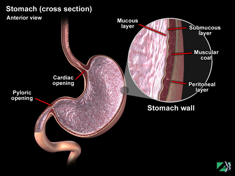
The lower end of the stomach, the end that joins the small intestine
Pyramid Fracture
A type of fracture of the maxilla, also known as Le Fort II fracture