Medical Glossary - Letter T
This medical glossary of terms beginning with the letter "T" contains the more common medical terms one might expect to encounter in a medical report or in hospital notes. The glossary is intended as a quick reference only; many of the terms are also referenced and illustrated in more detail in the medical libraries, to which you should refer for more detailed information.
Tachycardia
Rapid heartbeat
Tactile
Relating to the sense of touch
Talipes
A foot deformity, same as clubfoot. The foot may turn out or in
Talocalcaneal
Relating to the talus and the calcaneus
Talonavicular
Relating to the talus and the navicular, the joint formed by the talus and the navicular in the foot
Talus
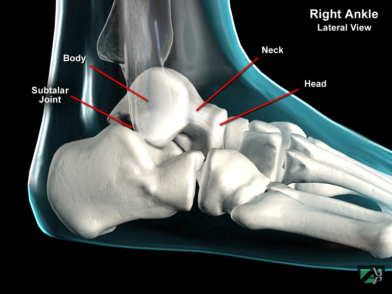
Although technically one of the tarsal bones of the foot, the primary significance of the talus is that it forms a vital part of the ankle joint. The talus articulates (connects) with the calcaneus below it and a socket above it formed by the lateral and medial malleoli. It also has an important weight bearing function as it transmits the weight of the body from the tibia to the foot, and also transmits forces from the foot to the leg. The talus consists of three parts, a head, neck and a body. The body of the talus is the part that fits into the socket created by the lateral and medial malleoli. The lower part of the body connects with the calcaneus to form what is known as the subtalar joint. The talar neck connects with the navicular bone while the head also forms part of the subtalar joint
Tarsal
Relating to the hind foot area of the foot, also known as tarsus
Tarsal Bones
The bones of the hind foot, they comprise the calcaneus (heel), talus, navicular, cuboid and three cuneiform bones
Tarsal Tunnel Syndrome
Tarsal tunnel syndrome is analogous to carpal tunnel syndrome in the wrist. It is caused by entrapment of the posterior tibial nerve or one of its branches, the medial and lateral plantar nerves. The tibial nerve enters the foot just below the medial malleolus and passes through a tunnel or canal formed by muscles and tendons. There are numerous causes of the syndrome but more often than not the cause is of an unknown origin (idiopathic), but there is a clear association with trauma. Compression of the nerve can result from fractures and dislocations of the tarsal bones
Tarsometatarsal
Refers to the area between the tarsal bones and the metatarsal bones of the foot
Tattooing
This is a plastic surgery procedure for cosmetic purposes. Tattooing involves injecting a pigment into the affected area of the skin to darken or lighten a blemish. Skin defects may also be injected to fill the defect
Temporal Bone
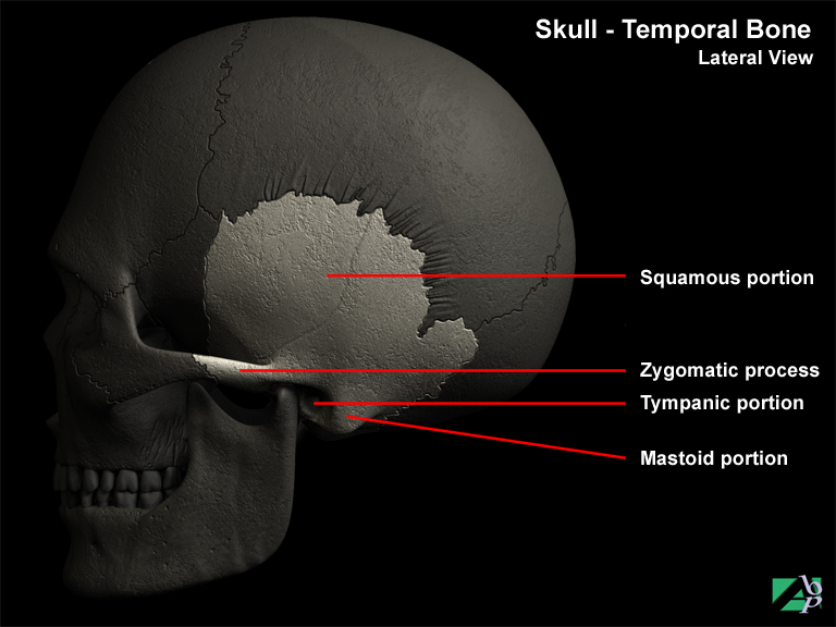
One of the bones of the skull, there are two, a left and a right, which form the floor and wall of the skull
Temporomandibular Joint
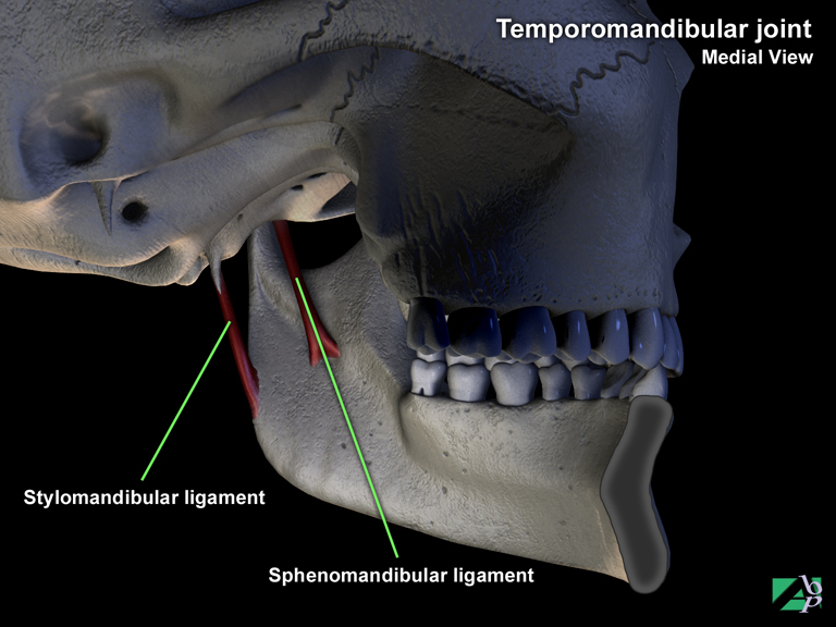
The joint formed by the mandibula fossa, a small depression in the temporal bone and condyle of the mandible
Temporomandibular Joint Dysfunction Syndrome
Temporomandibular joint dysfunction syndrome or TMJ describes an abnormal disorder of the temporomandibular joint. The disorder may involve the articular structures of the joint or the muscles and ligaments that support the joint movement. Symptoms include headaches, jaw pain, limited jaw opening, and clicking or popping of the jaw on jaw opening. Numerous causes exist for the disorder including malocclusion, bruxism (teeth grinding), stress, jaw injury, osteoarthritis, and whiplash is also sometimes blamed for the disorder
Tendinitis
Inflammation of a tendon, overuse is the most common cause of tendinitis
Tendon
A tendon is a tough fibrous tissue band that connects a muscle to the periosteum of a bone, which is the outer layer of the bone
Tendon Pulley
Tendon pulley refers to fibrous tissue called annular bands or pulleys that are part of tendon sheaths. This is known as the tendon pulley system. They attach to bones and hold tendons close to the bones over which they pass. The pulleys run diagonally across the tendon sheath
Tenonectomy
A tenonectomy is the surgical removal of part of a tendon. Tenonectomy is usually performed to shorten a tendon giving the muscle more tension and power
Tenosynovitis
Inflammation of a tendon sheath, sometimes known as tendosynovitis
Tenotomy
Tenotomy is the surgical incision (cutting) of a tendon
Tensor Fascia Lata
A muscle of the upper leg that originates from the iliac crest of the pelvis and inserts onto the lateral condyle of the tibia. It maintains knee extension and assists the gluteus maximus and abducts the hip. The superior gluteal nerve innervates it
Tentorium Cerebelli
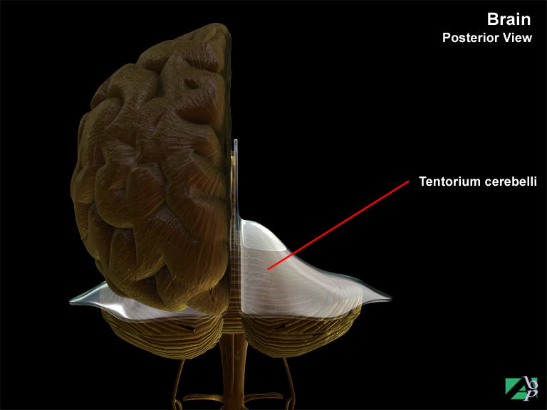
The tentorium cerebelli or just tentorium is an important structure because of its location, and anatomical relationships. The tentorium supports the occipital lobes of the cerebrum. It is attached at the sides and behind to the inner surface of the occipital bone, and in front to the petrous temporal bone. The anterior border of the tentorium is a free edge, concave in shape, and surrounding a large midline opening, the incisura tentorii or notch. The brainstem passes through this opening that is important in terms of trauma. Sudden violent movement of the brain from an impact to the head may throw the brain against the sharp free edge of the incisura, causing brain contusion or laceration. Additionally, when there is swelling or edema of the brain with increased intracranial pressure, the under side of the medial portions of the hemispheres may herniate (transtentorial herniation) through the opening of the tentorium, compressing the brain stem
Teres Major
A shoulder muscle. It originates from the scapula and attaches to the proximal end of the humerus (upper end). The muscle assists adduction and rotation of the arm
Teres Minor
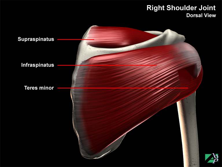
A shoulder muscle. It originates from the scapula and attaches to the greater tubercle of the humerus. It assists rotation and adduction of the arm
TFCC
An acronym for triangular fibrocartilaginous cartilage complex. The complex or structure comprises an articular disc (the triangular fibrocartilage), the ulnocarpal ligament, and the dorsal and volar radioulnar ligaments. It originates from the medial (inner) border of the distal radius and inserts onto the base of the ulna styloid and onto the lunate and triquetrum. The cartilage is thicker on the ulna side than on the radius side.
Thalamus
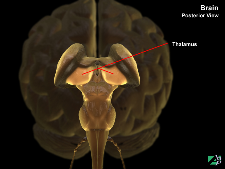
A part of the brain consisting of grey matter, located at the base of the brain. It is a relay center for sensory impulses being transmitted from areas of the body to the cerebral cortex for evaluation
Thecal
Pertains to the sheath of a tendon
Thenar
The bulge on the palm of the hand below the thumb, sometimes referred to as the thenar eminence
Thermocauterization
Thermocauterization is the destruction of a lesion by means of heat for example, an electrically heated wire
Thermokeratoplasty
This is a relatively new procedure in which a high temperature laser heats the cornea in 16 spots around its periphery. This heating shrinks the collagen causing the center of the cornea to steepen. The procedure is performed to correct refractive errors e.g., short sightedness and long sightedness
Theta Waves
A type of brain wave occurring mainly in children. In adults, theta waves may signify emotional stress
Thigh
Describes the region of the lower extremity between the knee and the hip
Third Ventricle
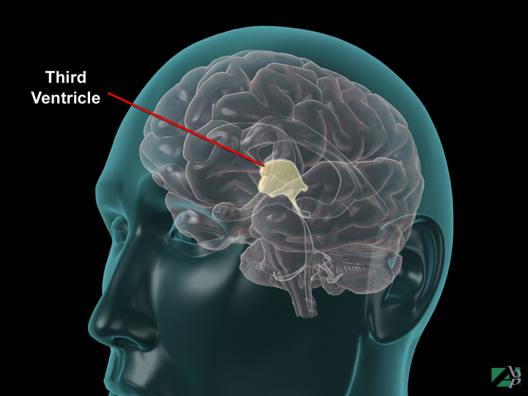
A chamber within the brain situated between the thalamus and hypothalamus, one of four such chambers within the brain where cerebrospinal fluid is produced
Thomas Splint
A traction device used mainly for fractures of the humerus and femur. It comprises a ring and two arms joined at the end by a crosspiece
Thoracentesis
An operation where the chest wall is punctured with a needle, usually to withdraw fluid
Thoracic
Relating to the chest area
Thoracic Aorta
The thoracic aorta is a branch of the aorta, which extends through the chest. The branches of the aorta supply blood to the chest wall, the thoracic organs and the upper part of the abdominal wall
Thoracic Cage
The thoracic cage is the rib cage, formed by the 12-paired ribs, the sternum and at the rear by the 12 thoracic vertebrae
Thoracic Cavity
The area within the chest containing the lungs, heart etc
Thoracic Outlet Syndrome
This describes a set of symptoms arising from compression of the neurovascular bundle in the shoulder. This neurovascular bundle consists of the brachial plexus, the major nerve plexus of the upper extremity and subclavian artery
Thoracolumbar
Relating to the thoracic and lumber regions of the spine
Thoracostomy
An operation to create a hole in the chest wall to drain fluid from the chest
Thoracotomy
An operation whereby an incision is made into the chest to open the chest and expose the thoracic contents
Thrombectomy
A surgical procedure where a surgical incision is made into a blood vessel to remove a thrombus (a clot)
Thrombophlebitis
Thrombophlebitis is blood clotting within a vein. Thrombophlebitis can result from numerous causes including prolonged bed rest, surgery, childbirth and use of oral contraceptives and from trauma
Thrombosis
The development of a blood clot, which is referred to as a thrombus
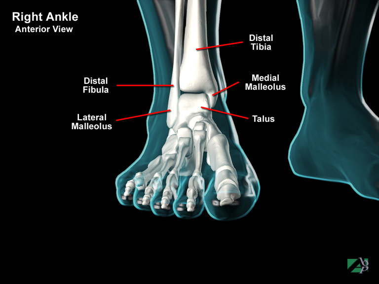
Tibia
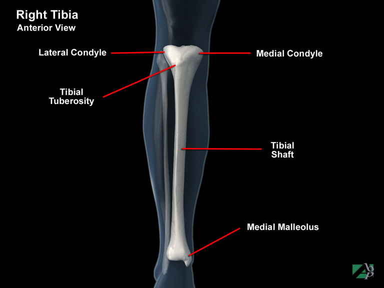
This is often referred to as the shinbone. The tibia can be divided into a proximal (knee) end, a shaft and a medial (ankle) end. At the proximal end, the tibia connects to the femur. Two concave surfaces at the proximal end called the medial and lateral condyles connect with the condyles of the femur. Just below and between the two condyles is a bony projection called the tuberosity. Distally at the ankle, the tibia has a flared knob like projection called the medial malleolus. The medial malleolus and the lateral malleolus (a similar knob like projection of the fibula), form a socket, which the talus of the foot fits into. The tibia and fibula are bound together top and bottom and along their entire course by a membrane called the interosseus membrane (a similar connection exists between the radius and the ulna)
Tibial Collateral Ligament
A ligament of the knee, which supports the inside of the knee. It extends from the femoral medial epicondyle and attaches to the medial condyle of the tibia
Tibial Nerve
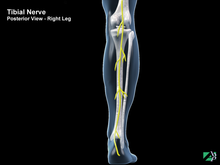
A peripheral nerve at the back of the knee. It is a continuation of the sciatic nerve and extends from the knee down the back of the leg to the foot. It innervates the calf muscles and provides sensation to the skin of the back of the leg and parts of the foot
Tibialis Anterior
A muscle of the leg. It extends from the knee down the front of the leg and attaches to the 1st metatarsal and 1st cuneiform bones in the foot. It assists inversion and dorsiflexion of the foot
Tibialis Posterior
A muscle of the leg. It extends from the back of the knee, down the back of the leg and attaches to the planter (underside) surface of the foot. It assists plantar flexion and inversion of the foot
Tinnitus
Tinnitus describes a ringing or buzzing in the ears. There are two forms: objective tinnitus and subjective tinnitus. Objective tinnitus is a form of tinnitus that can be heard by another person, a medical examiner for example. It is usually due to an abnormality of the blood vessels of the head and neck. The sound produced is due to the forceful passage of blood through constricted blood vessels. Subjective tinnitus is the form that can only be heard by the patient himself or herself. Tinnitus due to trauma is most often the result of an assault to the auditory nerve and/or inner ear mechanisms through head trauma or blast injuries and is often seen along with vertigo and loss of hearing
Tongue
The tongue is an organ with the principle functions of aiding speech and digestion of food. The tongue comprises a mass of striated (striped) muscle covered by a mucous membrane. The under surface of the tongue is connected to the floor of the mouth by the lingual frenulum. A septum of alveolar tissue provides a partial separation of the tongue into symmetrical halves. The groove in the midline on its dorsum is called the median sulcus. Three major extrinsic muscles control movements of the tongue: genioglossus, styloglossus and hyoglossus. The tongue has two parts, the anterior two-thirds which lies within the oral cavity proper and the posterior one-third, which lies within the pharynx. A V-shaped groove, the sulcus terminalis, separates the anterior portion in the mouth from the posterior pharyngeal portion. The upper surface of the tongue has numerous small elevations called the papillae, this roughened surface aids in handling food. These papillae also contain the taste buds. On the under surface of the tongue, the thin mucosa is reflected onto the adjacent gums and the floor of the mouth where it forms an elevated vertical fold in the midline, which is called the frenulum
Tooth Implant
Tooth implantation involves replacing a missing tooth with an implanted tooth. This is a staged procedure. First a metal or carbon post is surgically implanted into the bone. Once the bone has grown around the implant a porcelain crown is constructed around the post
Topical
Local, on the surface of the skin as in topical antiseptic or topical anaesthesia
Torticollis
A contraction of the neck muscles causing the head to tilt to the side, usually due to contraction of the sternocleidomastoid muscle
Toxaemia
Blood poisoning, a condition marked by the presence of a toxic substance in the blood
Trachea
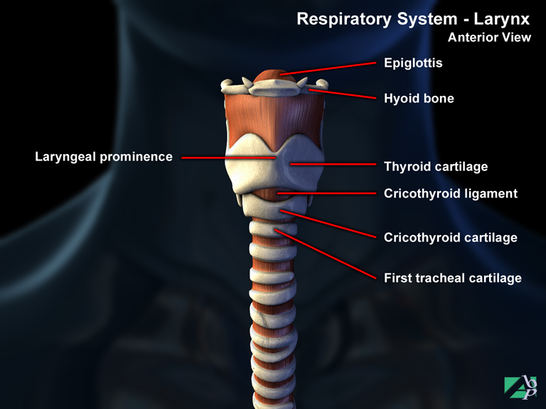
The trachea, or windpipe, connects with the larynx above to the bronchi of the lungs. It lies behind the esophagus. Its purpose is to conduct air to and from the lungs. It is a rigid tube composed of tough membrane supported by a series of circular cartilages to prevent it from collapsing. The inner surface of the trachea is lined with mucus secreting goblet cells and contains hairlike cells (cilia) that act to capture and expel foreign particles caught in the trachea. As the trachea enters the thorax it divides to join the two bronchi of the lungs
Tracheal
Relating to the trachea
Tracheobronchial
Relating to the trachea and the bronchus
Tracheostomy
A tracheostomy (also known as a tracheotomy) is a surgical procedure in which an opening is made in the windpipe (trachea). A tube is usually inserted into the opening to allow for removal of secretions from the lungs. It is often an emergency procedure performed because of a blockage of the airway, or an inhalation injury. The procedure involves making an incision into the neck and exposing the cartilaginous rings that form the outer wall of the trachea, two of the rings are cut and the tube inserted
Traction
Traction means to pull or draw on. Traction is often employed in the management of fractures; after the fracture is reduced traction is applied to maintain the position of the reduced fracture
Transcervical Fracture
A fracture occurring through the neck of a bone
Transected
Cut right through, a term often used to describe an injury to an artery or peripheral nerve
Transureteroureterostomy
Transureteroureterostomy, or transureteroureteral anastomosis is a surgical procedure in which one ureter (tube that carries urine from the kidney to the urinary bladder) is joined to its collateral ureter, so that the urine from the kidney empties into the bladder from one ureter. This procedure may be necessary if part of a ureter was so badly damaged that it could not be repaired
Transverse
Across
Transverse Acetabular Ligament
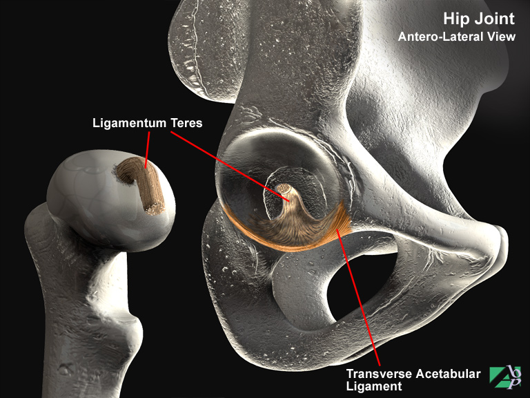
A ligament that supports the hip joint
Transverse Humeral Ligament
A shoulder ligament, it helps support the shoulder
Transversospinalis Multifidus
A long muscle of the back extending along the vertebral column. It extends the spine
Trapezium
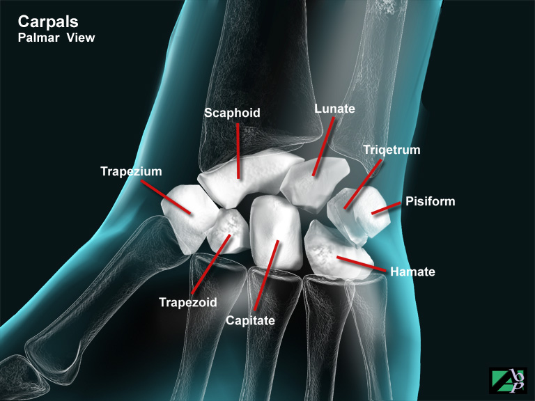
One of the carpal bones of the wrist, sometimes called the greater multangular bone
Trapezius
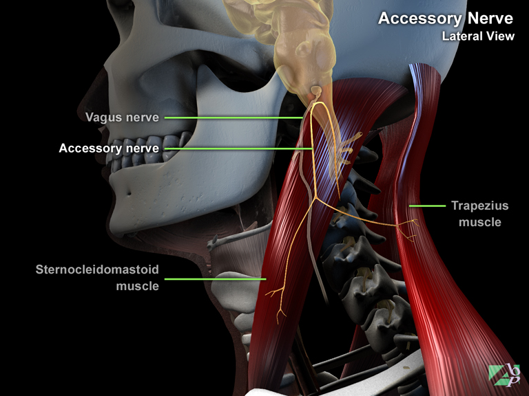
A large muscle of the back originating from the occipital bone at the back of the skull and inserting onto the clavicle and scapula. It assists movement of the scapula
Trapezoid
One of the carpal bones of the wrist, also known as the lesser multangular
Traumatic Shock
Traumatic shock is a state of severe circulatory compromise bought about by neurogenic and hypovolemic shock as a result of emotional stimuli to injury and pain, and severe blood loss. Shock brought about by emotional stimuli is referred to as neurogenic shock and is generally short-lived. Shock as a result of blood loss is known as hypovolemic shock. The combination of these two, unless checked and promptly treated result in circulatory collapse and death, although sufficient blood loss on its own will also result in circulatory collapse
Triangularis
A facial muscle located at the corner of the mouth, it acts to depress the corner of the mouth
Triceps Brachii
A muscle at the back of the upper arm. It has three heads, which originate from the scapula and humerus; it courses down the back of the upper arm and attaches to the ulna. It assists elbow extension
Trigeminal Neuralgia
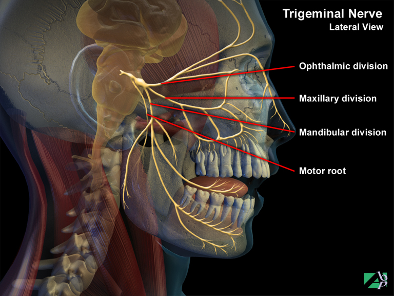
Facial pain resulting from injury or some disorder of the trigeminal nerve or its branches
Trimalleolar Fracture
A fracture of the ankle, involving fractures of both the lateral and medial malleolus and the lip (posterior) part of the tibia
Triquetrum
A carpal bone of the wrist. Also known as the triquetrium or triquetral bone.
Trochanter
The trochanters called the greater and lesser trochanters are two bony protuberances below the neck of the femur. They serve as points of attachment for major muscles of the thigh
Trochlea
Part of the distal humerus. The trochlea is the point of articulation with the semilunar notch of the ulna at the elbow
Trophic Ulcer
An ulcer on the skin that develops due to lack of blood supply to the area
Tubercle
A rounded outgrowth on a bone, such as found on the radius, humerus and tibia
Tympanic Cavity
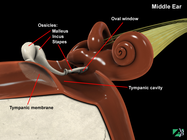
A cavity in the temporal bone of the skull containing the ear ossicles
Tympanic Membrane
The eardrum
Tympanoplasty
Tympanoplasty is plastic surgery for dysfunction of the tympanic membrane, the eardrum and the ossicular chain. Tympanoplasty is often classified as being type I, II, III, IV and type V. Type II tympanoplasty is performed for tympanic membrane perforations with erosion of the malleus (one of the three ossicular bones that transmit vibrations to the inner ear). Type III tympanoplasty is indicated when the lateral ossicles (malleus and incus) are destroyed but the stapes is intact, it involves placing a graft onto the stapes. Type IV tympanoplasty is indicated for ossicular destruction and involves removing part or all of the stapes arch and placing a graft into or around a stapes footplate. Type V tympanoplasty is indicated when the footplate of the stapes is fixed
Tympanostomy Tube
A tympanostomy tube is a tube placed through the tympanic membrane (eardrum) to drain fluid or pus in the inner ear
Tympanosympathectomy
Tympanosympathectomy is a surgical procedure where certain nerves are removed to relieve the symptoms of intractable tinnitus. The nerves removed are the tympanic plexus nerves, which are a bunch of nerves situated in the middle ear. They are comprised of the branches of the facial nerve and branches from the internal ceratoid plexus