Medical Glossary - Letter F
This medical glossary of terms beginning with the letter "F" contains the more common medical terms one might expect to encounter in a medical report or in hospital notes. The glossary is intended as a quick reference only; many of the terms are also referenced and illustrated in more detail in the medical libraries, to which you should refer for more detailed information.
Facet Joint Block
Facet joints are small articulating joints in the vertebra, they are also known as zygapophyseal joint. A facet joint block is the injection into the joint of either an anesthetic for pain relief or corticosteroid to reduce inflammation. The injection is done by needle under the guidance of a fluoroscope
Facetectomy
The removal of an articular facet of a vertebra
Facial Bones
The facial bones are bones of the skull that form the skeletal framework of the face, they are: maxilla, mandible, zygoma, nasal and orbit
Falx Cerebri
The falx cerebri is a partition formed by the dura mater (the outer layer of the meningeal covering of the brain) formed by an infolding or reduplication of the inner layer of dura in the midline, above, and extends downward separating the two cerebral hemispheres
Fascia
Fascia is a layer of tough tissue that covers, separates and supports body structures. It is found all over the body under the skin. Additionally it covers muscles and internal organs such as the kidneys and liver
Fasciectomy
A fasciectomy is the surgical excision of a piece of fascia, which is tough tissue beneath the skin
Fasciotomy
A fasciotomy is the surgical incision of fascia, which is the tissue beneath the skin and that surrounds organs, it provides support and firmness to the body and organs. Most often fasciotomy is performed for a condition known as compartment syndrome, in which structures such as blood vessels and nerves become compressed within confined areas of fascia resulting in neurovascular compromise. In the limbs, this is a limb threatening condition requiring emergency fasciotomy, which relieves the pressure within the fascia compartment. The forearm and calf area of the leg are the areas of the body where fasciotomy is most often performed
Fat Embolism
An embolism is a blockage of the circulatory system. The embolism may be comprised of air, fat or blood clot, or other material. Fat embolism describes such a blockage when the occluding agent is fat. Almost all fractures of the long bones and most notably femoral fractures will produce globules of fat that enter the circulation. Fat embolisms are either pulmonary or cerebral i.e., they lodge in either the lungs or they are systemic and can affect any tissues but most significantly the brain. Pulmonary fat embolism is the more common type of fat embolism. Fat globules enter the bloodstream within a very short time after fracture and perhaps even within seconds of the trauma and lodge within the lungs. Small accumulations do not impair respiratory function but larger accumulations do. Manipulation of the fracture in the reduction stage also contributes to the release of further fat into the blood stream
Femoral Artery
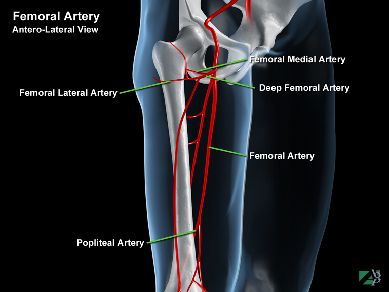
The femoral artery passes into the leg at an area known as the femoral triangle, a triangular area in the uppermost part of the inner thigh. At this point it gives off two branches, the deep femoral artery and the femoral lateral and medial circumflex branches. The deep femoral artery branches to the back of the thigh where it supplies the hamstring muscles. The lateral and medial femoral circumflex branches supply the muscles around the upper end of the femur. The femoral artery then continues down the back of the leg to the knee where it becomes the popliteal artery
Femoral Nerve
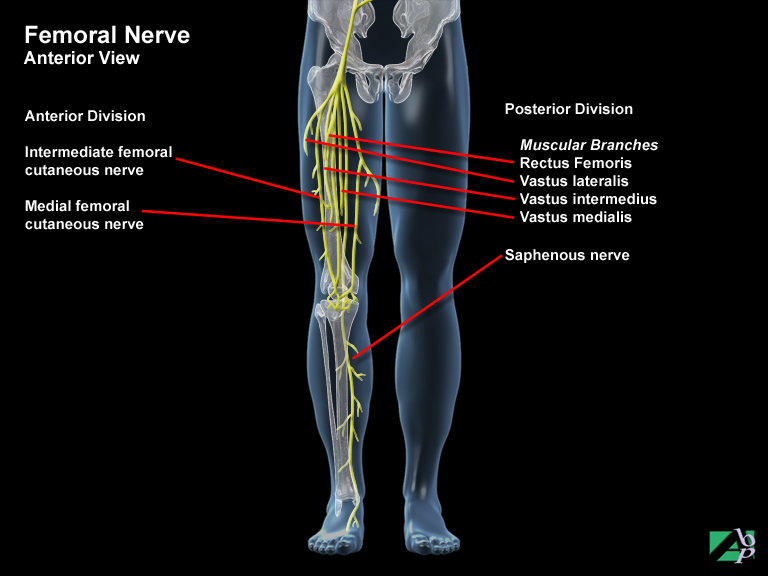
The femoral nerve is a mixed nerve (motor and sensory) and emerges from the abdomen and passes down the front of the thigh to the knee where it becomes the saphenous nerve. In the thigh it provides motor impulses to activate the muscles that flex the thigh and help extend the knee. The muscles include the iliacus, psoas major, pectineus, rectus femoris and sartorius muscles and vastus lateralis and vastus medialis. The femoral nerve also supplies sensation to the front and inside of the thigh
Femur
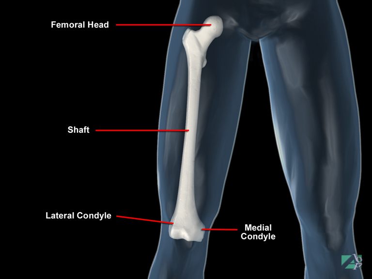
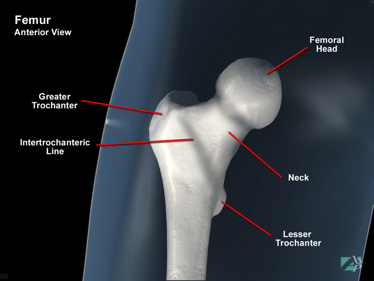
This is the largest bone in the body. It extends from the hip to the knee and is divided anatomically into three parts, a proximal femur, a shaft and a distal femur. The proximal femur begins at the femoral head, which forms a joint with the acetabulum to become the hip. Just below the head is a narrowed region called the femoral neck, just below the neck at the beginning of the shaft, are two bony protuberances called the greater and lesser trochanters. At the lower end of the shaft, the distal femur, there are two prominences called the medial and lateral condyles. These condyles join with the upper ends of the tibia and fibula to form the knee joint
Fenestration
Fenestration describes a surgical procedure where an artificial opening, like a window is made into the labyrinth of the ear. The labyrinth is a series of cavities within the temporal bone of the skull, which contains the structures of the inner ear, the cochlea, the vestibule and the semicircular canal
Fiber-Optic Bronchoscopy
Fiberoptic bronchoscopy is an investigative procedure used to visualize the larynx, trachea and the bronchial air passages (bronchus). The procedure is performed using a bronchoscope, which is a long flexible tube with a viewing light at the end. A camera may or may not be attached to the other end. The bronchoscope is passed usually by the nose but may be passed through the mouth. Tissue biopsy may also be performed during the test
Fibrocartilage
Fibrous tissue within or supporting cartilage
Fibrositis
Inflammation of tissue, muscle fibers and / or other layers of fascia. The symptoms are generally aching and stiffness of the muscles affected
Fibula
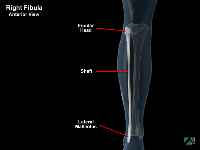
The fibula is a bone of the lower leg. The main function of the fibula is for muscle attachment; it provides little in the way of support to the leg. The fibula comprises a proximal end, a shaft and a distal end. The upper or proximal end has a head, which connects with the proximal end of the tibia (but not with the femur). Below the head is a narrower section referred to as the neck. At the distal (ankle) end it forms a knob like projection called the lateral malleolus, which connects with the talus of the foot and forms part of the ankle joint
Fistula
A fistula is an abnormal opening or communication between two organs or structures of the body
Fixed Bridge
A fixed bridge may be used to replace a missing tooth or teeth. It involves an appliance with a false tooth or teeth that is held in place by being attached to an adjacent tooth, which is known as an abutment tooth. It requires the abutment teeth to be crowned in order to accept the bridge. Once the bridge is in place; the patient cannot remove it
Flail Chest
Flail chest is a condition that sometimes results from multiple rib fractures that occur at separate locations that result in free floating rib segments. It is a serious injury and one that severely compromises respiration and can be life threatening. It is often associated with contusions to the lungs and hemothorax and pneumothorax and lung collapse. When flail chest occurs, it reverses the normal mechanism of breathing. The injured area moves outward with inspiration, yet the uninjured area moves inward, conversely with expiration, the injured ribs force the chest outward while the uninjured part of the chest moves inward. The result of this is inefficient gas exchange within the lungs and oxygen is not transported into the blood sufficiently and lack of blood oxygen results
Flank
The flank refers to the side of the body, that part between the ribs and the hipbone
Fluoroscopy
Fluoroscopy, an imaging technique that projects an x-ray type picture onto a monitor
Flexion
Flexion is a term used to describe bending part of a limb, for example to grasp an object it requires flexing the fingers to grip the object. Flexion is the opposite movement to extension. In the example given, to release a grasped object it requires extension or straightening of the fingers. Muscles that allow flexion of a body part are known as flexor muscles and their tendinous attachments as flexor tendons
Flexor Carpi Radialis
A muscle originating from the medial epicondyle of the humerus at the elbow and extending down the forearm and inserting onto the base of the 2nd and 3rd metacarpals. It provides flexion and abduction of the wrist
Flexor Carpi Ulnaris
A muscle originating from the medial epicondyle of the humerus at the elbow and extending down the forearm and inserting onto the pisiform and hamate bones in the wrist and the base of the 5th metacarpal. It flexes and adducts the wrist. The ulnar nerve innervates it
Flexor Digiti Minimi Brevis of Foot
A muscle of the foot, originating from the base of the 5th metatarsal (behind the little toe) and inserting onto the metatarsophalangeal joint of the little toe. It flexes the metatarsophalangeal joint. The lateral plantar nerve innervates it
Flexor Digiti Minimi Brevis of Hand
A muscle of the hand originating from the hamate bone in the wrist and inserting onto the proximal phalanx of the little finger. The ulnar nerve innervates it. The muscle flexes the metacarpophalangeal joint of the little finger
Flexor Digitorum Brevis
A muscle in the foot originating from the tuberosity of the calcaneus (heel) and inserting onto the 2nd, 3rd, 4th and 5th toes. It flexes the four toes. The medial plantar nerve innervates it
Flexor Digitorum Longus
A muscle originating from the shaft of the tibia in the lower leg, which extends down the leg into the foot and inserts onto the 2nd, 3rd 4th and 5th toes. It flexes the toes and the foot at the ankle. The tibial nerve innervates it
Flexor Digitorum Profundus
A muscle originating in the forearm from the ulna, which extends down the forearm and inserts onto the distal phalanges of the index, middle, ring and little fingers. It flexes the distal interphalangeal joints and wrist. The median and ulnar nerves innervate it
Flexor Digitorum Superficialis
A muscle originating from the distal humerus at the elbow, which extends down the forearm and inserts onto the middle phalanges of the index, ring, middle and little fingers. It assists flexion of the proximal interphalangeal and metacarpophalangeal joints of these fingers and the wrist
Flexor Hallucis Brevis
A muscle of the foot, which originates from the cuboid and cuneiform bones of the foot and inserts onto the proximal phalanx of the big toe and the sesamoid bones. It flexes the metatarsophalangeal joint of the big toe. The medial plantar nerve innervates it
Flexor Hallucis Longus
A muscle that originates from the lower part of the fibula, extends into the foot and inserts onto the distal phalanx of the big toe. It flexes the distal phalanx of the big toe and assists flexion of the foot. The tibial nerve innervates it
Flexor Pollicis Brevis
A muscle of the hand that originates from the trapezium at the wrist and inserts onto the proximal phalanx of the thumb. It flexes the thumb at the metacarpophalangeal joint. The median nerve innervates it
Flexor Pollicis Longus
A muscle originating in the front of the forearm from the radius and extending down the forearm into the hand and inserting onto the distal phalanx of the thumb. It flexes the distal phalanx of the thumb
Foot drop
A state in which the individual is unable to extend the foot upwards towards the leg, it is due to a peripheral nerve disorder in which the muscles that extend the foot are paralyzed due to the nerve innervating them being dysfunctional
Foramen Magnum
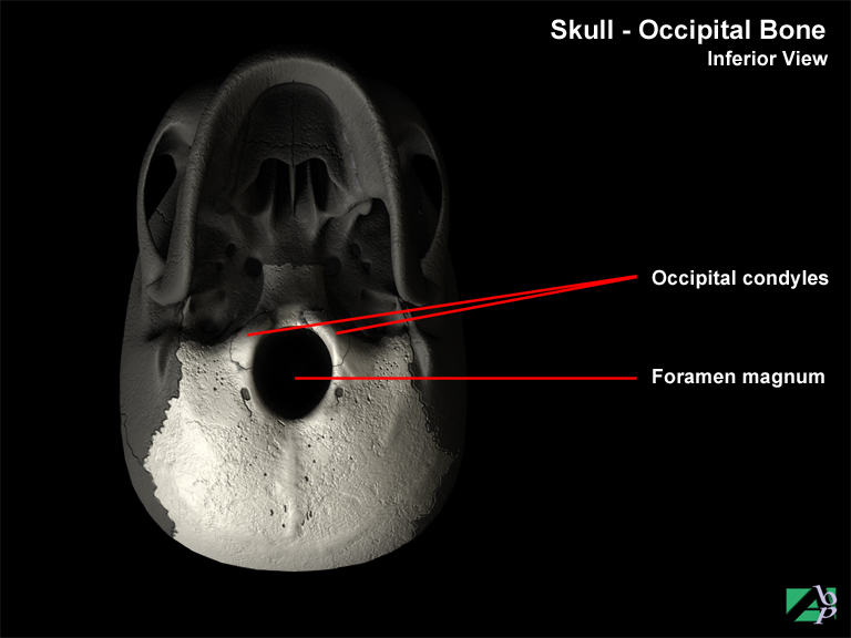
The foramen magnum is an opening in the base of the skull (in the occipital bone) that provides a passageway for the lower portion of the brainstem, as it becomes the upper portion of the spinal cord
Foraminotomy
Foraminotomy is a surgical procedure to widen the vertebral foramen, which is an opening in the vertebrae through which spinal nerves pass. It is usually performed to relieve pressure on spinal nerves
Fore Foot
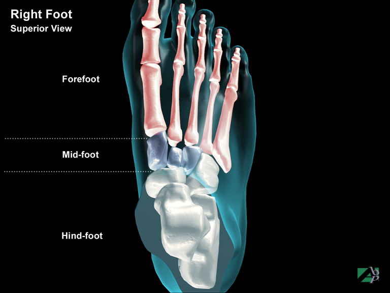
That part of the foot comprising the metatarsals and the toes
Fourth Ventricle
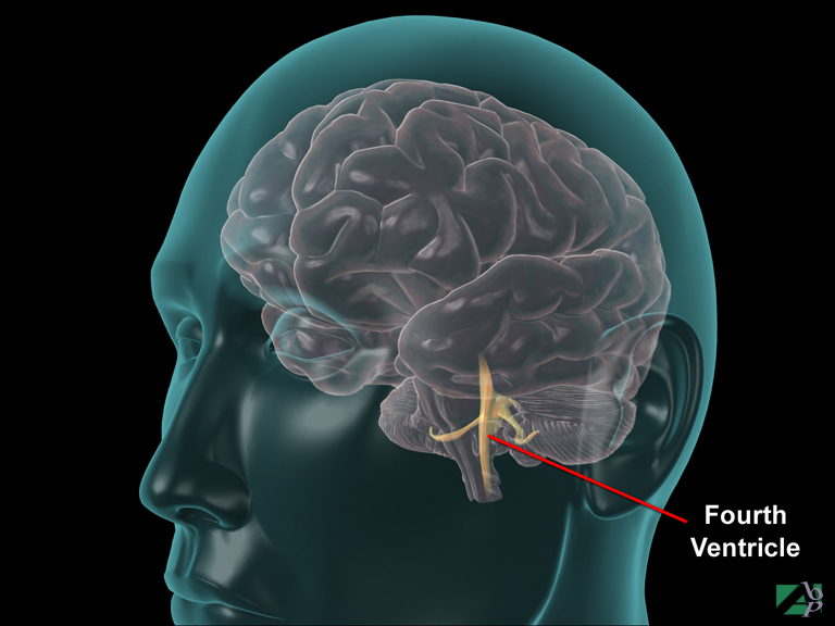
A chamber within the brain situated between the pons and the cerebellum. One of four such chambers where cerebrospinal fluid is produced
Fracture
A break in a bone
Fracture - Articular
A fracture of part of a bone (the end or head of a bone) that articulates with another bone, a fracture into a joint
Fracture - Avulsion
A fragment of bone torn away by a ligament or tendon of a muscle becoming detached from its insertion onto the bone. Severe sprains sometimes result in a fragment of bone becoming detached
Fracture - Barton's
A fracture of the distal radius at the radiocarpal joint (where the radius articulates with the carpal bones of the wrist)
Fracture - Basilar
A fracture of the base of the skull, the base of the skull is made up of the basal portion of the frontal bone, the ethmoid, the sphenoid and the basal portion of the occipital bone
Fracture - Bennett's
A fracture of the base of the first metacarpal of the hand, the first metacarpal bone is the metacarpal in line with the thumb
Fracture - Blowout
A fracture of the eye-socket (the orbit)
Fracture - Burst
A fracture in which the bone is fragmented into many pieces, it is sometimes used to describe a fracture of a vertebra
Fracture - Closed
A fracture not involving exposure of the fractured bone to the outside environment, i.e., with no associated protrusion of the bone or wound at the fracture site
Fracture - Colles'
A Colles' fracture is a fracture of the distal end of the radius with backward displacement
Fracture - Comminuted
A fracture in which the fracture bone is shattered into many pieces
Fracture - Complete
A fracture where the fracture line extends completely through the bone
Fracture - Compound
Same as open fracture, a fracture in which the fractured bone is exposed to the outside environment
Fracture - Compression
A fracture resulting from compression forces, the thoracic vertebrae are most vulnerable to fractures from compression forces
Fracture - Condylar
A fracture through a condyle of a bone, a condyle is a bony prominence found on some long bones such as the tibia and humerus
Fracture - Contrecoup
A skull fracture that occurs distant from the point of impact, for example a blow to the forehead may result in a fracture at the back of the head
Fracture - Depressed
A fracture in which a piece of bone is forced inwards, particularly of the skull, or zygoma (cheekbone)
Fracture - Epiphyseal
A fracture of the end of the shaft of a bone where the epiphyseal plate is located, the epiphyseal plate is the growth area of a long bone. This type of fracture only affects children or adolescents, in adults the epiphyseal plate closes and the cartilage is replaced by normal bone
Fracture - Galeazzi
A fracture dislocation involving a fracture of the distal (wrist end) radius and a dislocation of the head of the ulna
Fracture - Greenstick
An incomplete fracture that occurs in children
Fracture - Impacted
A fracture where one end of a broken bone is impacted into the other broken end
Fracture - Intracapsular
A fracture to a bone within the joint capsule, same as intra-articular fracture
Fracture - Le Forte
Fracture of the maxilla. Maxilla fractures are classified as LeFort fractures of which there are three types: LeFort I- is the most frequent type. It is a transverse fracture occurring just above the level of the teeth in the alveolar process. This fracture results in the roof of the mouth (the hard palate) separating. LeFort II is referred to as a pyramid fracture, as the fracture lines resemble the shape of a pyramid. In this vertical fracture, the fracture block contains the frontal processes of the maxilla and the nasal bones and is often associated with a depressed or comminuted fracture of a zygoma. The fractures also commonly extend through the lacrimal bones and the floor of the orbit. LeFort III is the most complex of all facial fractures as it involves the entire middle third of the face. The fractures extend from the nasofrontal junction down the medial wall of the orbit, across the floor of the orbit and through the maxilla, ethmoid and sphenoid bones, with complete separation of the middle facial skeleton
Fracture - Linear
This term describes a fracture where the fracture line extends lengthwise along the bone
Fracture - Monteggia
A fracture dislocation of the forearm involving a fracture of the proximal ulna (near the elbow) and a dislocation of the radial head
Fracture - Open
Same as compound fracture, a fracture in which the fractured bone is exposed to the open environment
Fracture - Pertrochanteric
A fracture of the femur in which the fracture extends through the trochanter of the femur, which are bony prominences below the femoral neck
Fracture - Stellate
A fracture where the fracture line(s) are starlike in appearance, they most commonly occur in the skull
Fracture - Subcapital
A fracture occurring just below the head of a bone
Fracture - Teardrop
A fracture of the vertebral body, they are unstable fractures and are frequently associated with spinal cord lesions at the level of the fracture
Fracture - Transcervical
A fracture occurring through the neck of a bone
Free Skin Graft
A free skin graft is a piece of skin completely detached from one site and used on another location. This is the opposite to another type of skin graft called a pedicle graft, where part of the graft is left attached in its original position, while the remainder is used as a graft nearby
Frenulum
The mucous membrane that connects the lip to the gum and the tongue to the floor of the mouth
Frontal Bone
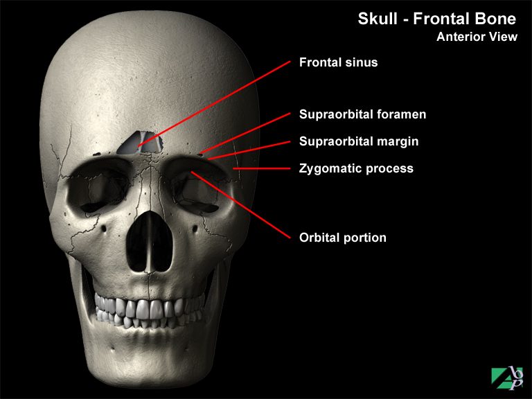
The frontal bone forms the anterior roof of the skull, the forehead, the roof of the nasal cavity and the superior arch of the orbits containing the eyeballs. The prominent bony ridge over the orbit (the eyebrows) is known as the supraorbital margin. Foramina along this ridge called supraorbital foramina, allow passage of small nerves and vessels. The frontal bone also contains a frontal sinus, which connects the nasal cavity, which acts as a sound chamber for voice resonance
Frontal Lobe
The frontal lobe is the anterior (front) portion of each cerebral hemisphere. The frontal lobes functions include initiating voluntary motor impulses for the movement of skeletal muscles, analyzing sensory experiences and providing responses relating to personality. The frontal lobes also involve responses related to memory, emotions, reasoning, judgement and planning and verbal communication. A highly specialized area of the frontal lobe is the motor speech area, known as Broca's area. Generally located in the left hemisphere, mental activity in Broca's area causes selective stimulation of motor impulses in motor centers elsewhere in the frontal lobe, which in turn cause coordinated skeletal muscle movement in the pharynx and larynx. At the same time, motor impulses are sent to the respiratory muscles to regulate air movement across the vocal cords. This combined muscular stimulation translates thought patterns into speech
Frontalis
A muscle lying over the frontal bone of the skull. It inserts onto the soft-tissue of the eyebrow and serves to elevate the eyebrow and wrinkle the forehead
Frozen Shoulder
Frozen shoulder is another term for adhesive capsulitis. Adhesive Capsulitis is a disorder that affects the shoulder only and its cause is unknown, though it has been associated with trauma. In some cases it is associated with calcium deposits. The disorder is seen much more frequently in women than in men. The primary symptoms of adhesive capsulitis are stiffness and pain and in some cases loss of shoulder motion can significantly limit the individual's use of the shoulder and upper arm
Full-Thickness Skin Graft
A full-thickness skin graft refers to a graft comprising all layers of the skin but without the subcutaneous tissue lying below