Medical Glossary - Letter U
This medical glossary of terms beginning with the letter "U" contains the more common medical terms one might expect to encounter in a medical report or in hospital notes. The glossary is intended as a quick reference only; many of the terms are also referenced and illustrated in more detail in the medical libraries, to which you should refer for more detailed information.
Ulcer
A skin erosion
Ulna
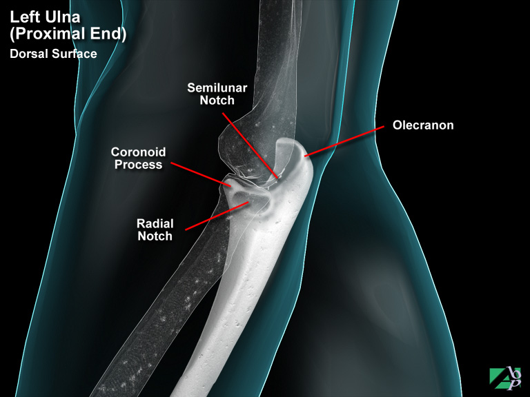
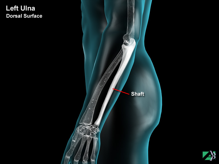
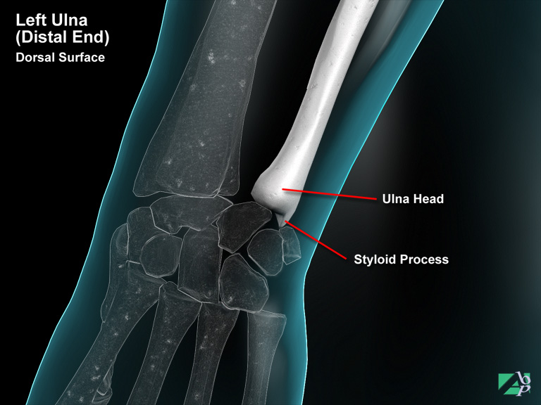
The ulna comprises a proximal (elbow) end, a shaft and a distal end (wrist). The proximal end is the elbow end, which connects with both the humerus and radius. The upper end of the ulna has a hook like spur called the olecranon. This is the point of the elbow. Below the olecranon is a bony lip known as the coronoid process. Between the olecranon and the coronoid process is a rounded cavity named the semilunar notch, which receives the trochlea of the humerus. On the outer side of the coronoid process is another rounded cavity called the radial notch, which receives the head of the radius. The shaft of the ulna is sometimes divided anatomically into thirds, an upper third, middle third and lower third. An important landmark on the shaft is a ridge called the interosseous crest. A membrane extends from this crest and attaches to the radius to bind the two bones together. The distal end, or wrist end of the ulna has a head and behind this a projection of bone called the styloid process, which is the point of attachment for the ulna collateral ligament of the wrist
Ulnar Artery
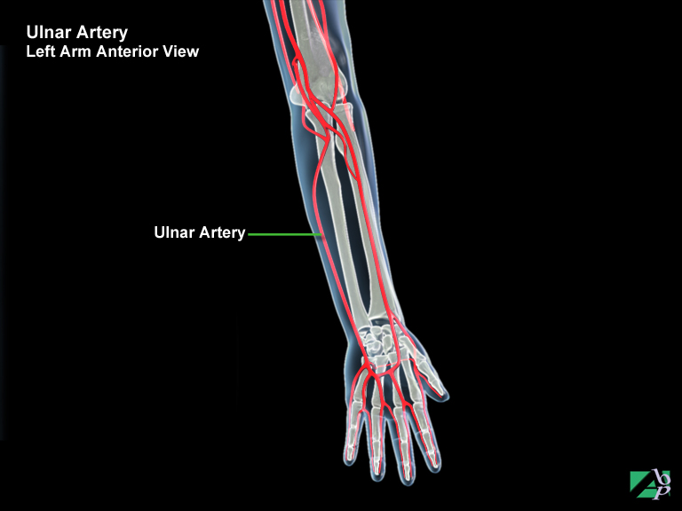
The ulnar artery is a continuation of a branch of the brachial artery and commences just below the elbow and descends down the ulna side of the forearm (the little finger side) to the wrist where it divides into superficial and palmar branches, which finally merge with the radial artery. At its point of origin below the elbow, other branches are given off to supply blood to the elbow. In its descent down to the wrist it supplies blood to the deeper structures of the forearm
Ulnar Collateral Ligament
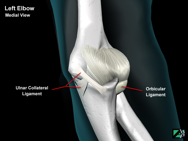
A ligament in the elbow, it helps support the elbow joint
Ulnar Nerve
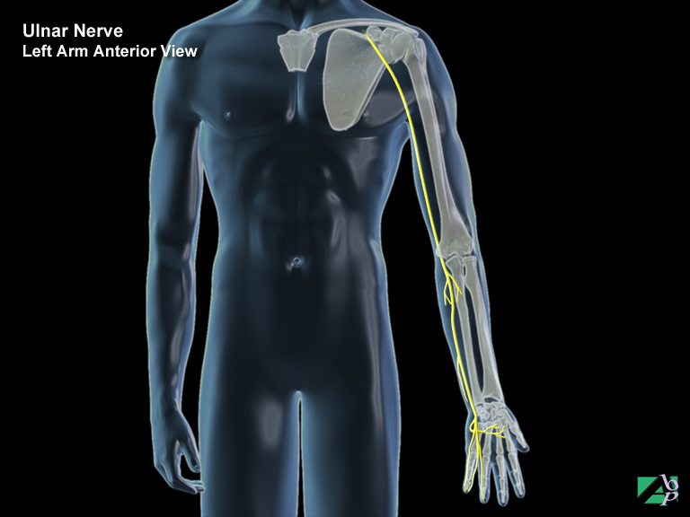
The ulnar nerve is a mixed nerve (motor and sensory). It supplies sensation to the skin of the little finger, ring finger and part of the hand (ulna side, or the side of the little finger) and motor innervation to the flexor carpi ulnaris and the deep finger flexors. Its main trunk supplies the hypothenar muscles
Ultrasound
An ultrasound is a diagnostic procedure and is an image of the body or internal organs created with the use of high frequency sound waves. A hand held instrument that emits the sound waves is run over the body part to be scanned, the organs or structures reflect the sound waves, which the instrument then passes to a computer to analyse
Umbilical Hernia
A hernia occurring at the navel
Umbilical Region of Abdomen
The umbilical region contains the jejunum, ileum, some of the duodenum, colon, kidneys and the major blood vessels. It describes the location of the center of the abdomen
Umbilicus
The navel
Unilateral
Affecting one or one side of the body
Upper G.I. Series
An upper G.I. series is an x-ray of the upper gastrointestinal tract made up of the esophagus, stomach and duodenum. It is essentially the same as a barium swallow
Ureter
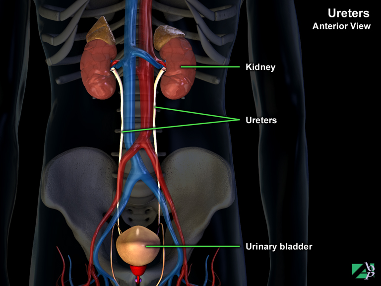
The paired ureters (one for each kidney) are tubular organs approximately 25 centimeters in length. Their function is to transport the urine and other waste matter formed by the kidneys to the urinary bladder
Ureteroileostomy
Ureteroileostomy (a form of urinary diversion) is the surgical joining of the ureters to a detached loop of the ileum, which is sutured to the abdominal wall to act as a stoma (artificial opening between two organs) and is used to drain urine from the body. The procedure may also be known as an ileal conduit or a continent ureterostomy
Ureteroneocystostomy
Ureteroneocystostomy is the surgical formation of a new opening between a ureter (or both ureters) and the bladder. It is performed when the normal connection of the ureter and bladder is non-functional or destroyed
Ureteroscopy
Ureteroscopy can be both a diagnostic procedure and minimally invasive surgical technique. It is performed using an endoscope, a small flexible fiberoptic tube, which is passed into the ureter, the tube that conveys urine from the kidney to the bladder. As a diagnostic procedure it allows visualization of the interior of the ureter. As a surgical technique it could for example be used to remove obstructions of the ureter, such as a stone
Ureterotomy
Ureterotomy is the surgical incision of the ureter, the tube that conveys urine from the kidney to the bladder. This may be necessary due to constriction or obstruction of the ureter following a traumatic injury
Urethra
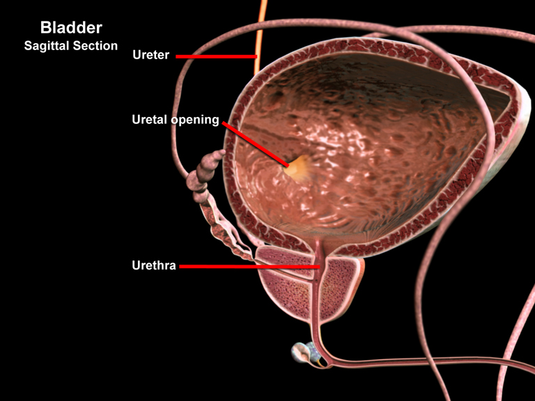
The urethra is a tubular organ that conveys the urine from the bladder to the outside. The size and function of the urethra differs between males and females. In females it is a simple tube approximately four centimeters in length and serves only to drain the urine from the body. In males, the urethra also plays a reproductive role. The male urethra is also much longer measuring approximately 20 centimeters in length. The male urethra is divided into three sections: the prostatic urethra that passes through the prostate, the membranous urethra and the penile urethra. The prostatic urethra receives drainage from the prostate gland and also ejaculatory ducts from the reproductive system
Urethral Meatus
The external opening of the urethra
Urethral Stricture
An abnormal narrowing or blockage of the urethra, it is a relatively common complication of males with urethral injuries
Urinary Diversion
Temporary or permanent urinary diversion may be required for injuries to the kidney or bladder or following surgical repair to the bladder or ureters (tubes that carry urine from the kidney to the bladder). The diversion is either cutaneous (through the abdominal wall) or into the intestine, which is referred to as an internal diversion