Medical Glossary - Letter M
This medical glossary of terms beginning with the letter "M" contains the more common medical terms one might expect to encounter in a medical report or in hospital notes. The glossary is intended as a quick reference only; many of the terms are also referenced and illustrated in more detail in the medical libraries, to which you should refer for more detailed information.
Macerated
This term describes a state of skin being raw and tender
Magnetic Resonance Imaging (MRI)
MRI is a radiological diagnostic procedure that utilizes a magnetic field and radio waves to project detailed images of the human body
Major Surgery
Any operation involving major body organs or involving significant blood loss or having potential life threatening implications
Malabsorption
This term describes abnormal absorption of nutrients from the digestive tract, particularly the small intestine
Maladie (Malady)
A term used to describe an illness or disorder
Maladjustment
An inability to adjust to circumstances in an adequate fashion
Malar
Another name for the zygoma (cheekbone)
Maldevelopment
Abnormal development
Maldigestion
Abnormal digestion
Maleruption
Teeth erupting in an abnormal position
Malformed
An abnormality, an abnormal development of a body structure
Malleolus
The rounded bony protuberances on the distal ends of the fibula and tibia, the one on the fibula is called the lateral malleolus and the one on the tibia is called the medial malleolus. They form part of the ankle joint
Mallet Finger
An extension deformity of a finger involving the extensor tendon of the terminal phalanx, usually as a result of the finger being forcibly bent during active extension. The index and middle fingers are the most affected digits
Malocclusion
An abnormal bite, the upper and lower teeth do not meet properly
Malunion
The term is used to describe a fracture, which has united but not in proper anatomical union
Mandible
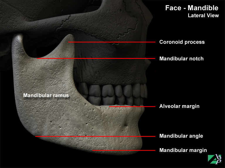
The lower jaw bone. It is horseshoe in shape with two U shaped ends that are known as the ramus. The rest of the mandible (the chin) is referred to as the body. It consists of two halves that unite at the chin. The ridge of bone that forms the upper border of the body is the alveolar process that houses the sockets for the roots of the lower teeth. Each ramus has a knoblike condyloid process, which fits into a depression in the temporal bone on the underside of the skull known as the temporomandibular joint. The ramus also has a pointed coronoid process for the attachment of the temporalis muscle. A depressed area between these two processes is the mandibular notch. The junction of the body and ramus of the mandible is called the angle. Two sets of foramina (openings) are found on the mandible: the mental foramen on the lateral side of the first molar tooth and the mandibular foramen on the medial surface of the ramus. The mental nerve and vessels pass through the mental foramen, and the inferior alveolar nerve and vessels are transmitted through the mandibular foramen
Mandibular
Relating to the mandible
Mandibular Nerve
The mandibular nerve is a branch of the trigeminal nerve (Cranial Nerve V). The mandibular branch innervates muscles that are used in chewing and provides sensation to areas of the tongue and mouth
Manubrium
The upper portion of the sternum
Marsupialisation
A surgical procedure performed on cysts. An incision is made into the cyst, which is then left open (like a pouch) to be filled with granulating tissue (tissue produced in the healing process of a wound).
Masseter Muscle
A facial muscle. Its point of origin is the cheekbone and its point of attachment is the mandible. It assists in raising the lower jaw
Mastication
This term describes the act of chewing food
Mastoid
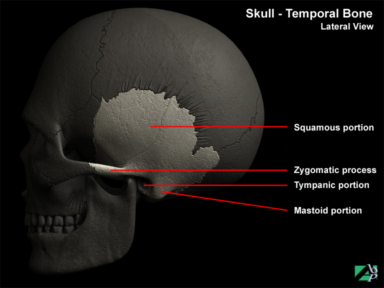
Part of the temporal bone of the skull, situated behind the ear
Mastoidectomy
Mastoidectomy is the surgical removal of the mastoid process and mastoid air cells of the temporal bone. There are variations of the procedure including what is referred to as simple mastoidectomy and radical mastoidectomy. Simple mastoidectomy involves only the removal of the pyramid air cells contained in the mastoid. Radical mastoidectomy involves removal of these air cells along with the ossicular chain (three small bones that transmit vibrations to the inner ear) and also part of the labyrinth bone of the inner ear
Maxilla
The upper jaw bone. There are two maxillae that unite in the center of the face and form the upper jaw bone. The lower border of the bone known as the alveolar process contains the sockets that hold the roots of the upper teeth. The upper part of the maxilla partly forms the floor of the orbits (eye sockets). The lower part forms part of the walls of the nose and the roof of the mouth. At the side near the nose a large space is located named the maxillary sinus or maxillary antrum
Maxillary
Relating to the maxilla
Maxillary Nerve
A branch of the trigeminal nerve (Cranial Nerve V). It provides sensation to the upper teeth, part of the nose, roof of the mouth and to part of the skin of the face
McMurray's Sign
Also known as McMurray's test. A test for meniscus injury, if the meniscus is injured a clicking sound may be heard when the knee is manipulated
Meatoplasty
An operation on the urethra
Mechanical Ventilation
Mechanical ventilation is a method for using machines to help a patient breathe when they are unable to breathe sufficiently on their own. Generally mechanical ventilation is used only for a couple of days in the acute phase. Mechanical ventilation may be necessary for head injury patients and patients with life threatening thoracic injuries or respiratory complications
Medial (Median)
A term used to describe the innermost part of a body structure, the part nearest or facing the middle of the body. For example the inner thigh is the medial aspect of the thigh, whereas the outer side of the thigh facing away from the midline is the lateral aspect
Medial Collateral Ligament
A ligament of the knee, it holds the medial meniscus in place. The deltoid ligament in the ankle is also sometimes known as the medial collateral ligament
Medial Condyle of Femur
The medial condyle of the femur is a bony prominence on the end of the femur, it articulates with the medial tibial plateau of the tibia
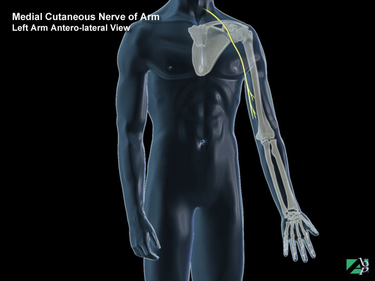
Medial Cutaneous Nerve
The medial cutaneous nerve is a sensory nerve that provides sensation to the skin of the medial side of the forearm (the side towards the body)
Medial Gastrocnemius Bursa
A bursa situated in the knee
Medial Malleolus
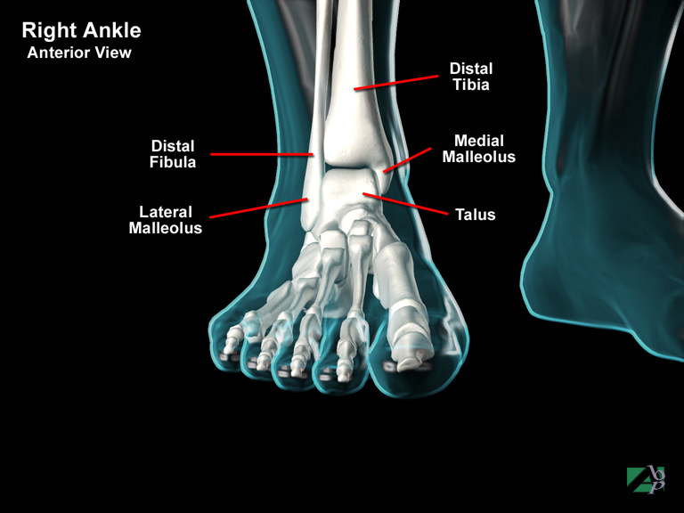
A bony knob on the distal (lower) end of the tibia, it forms part of the ankle joint
Medial Meniscus
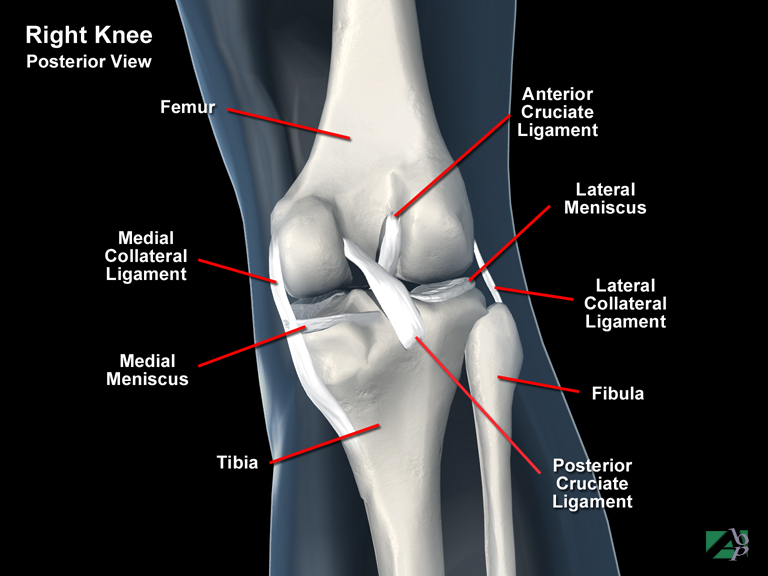
One of the two menisci of the knee, it is a crescent shaped cartilage that lies between the femur and the tibia. It assists load bearing, and acts as a shock absorber and lubricates the joint
Medial Plantar Artery
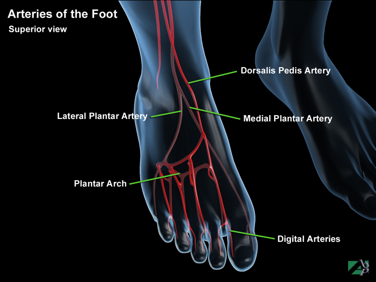
An artery that commences in the ankle and supplies the sole of the foot
Medial Plantar Nerve
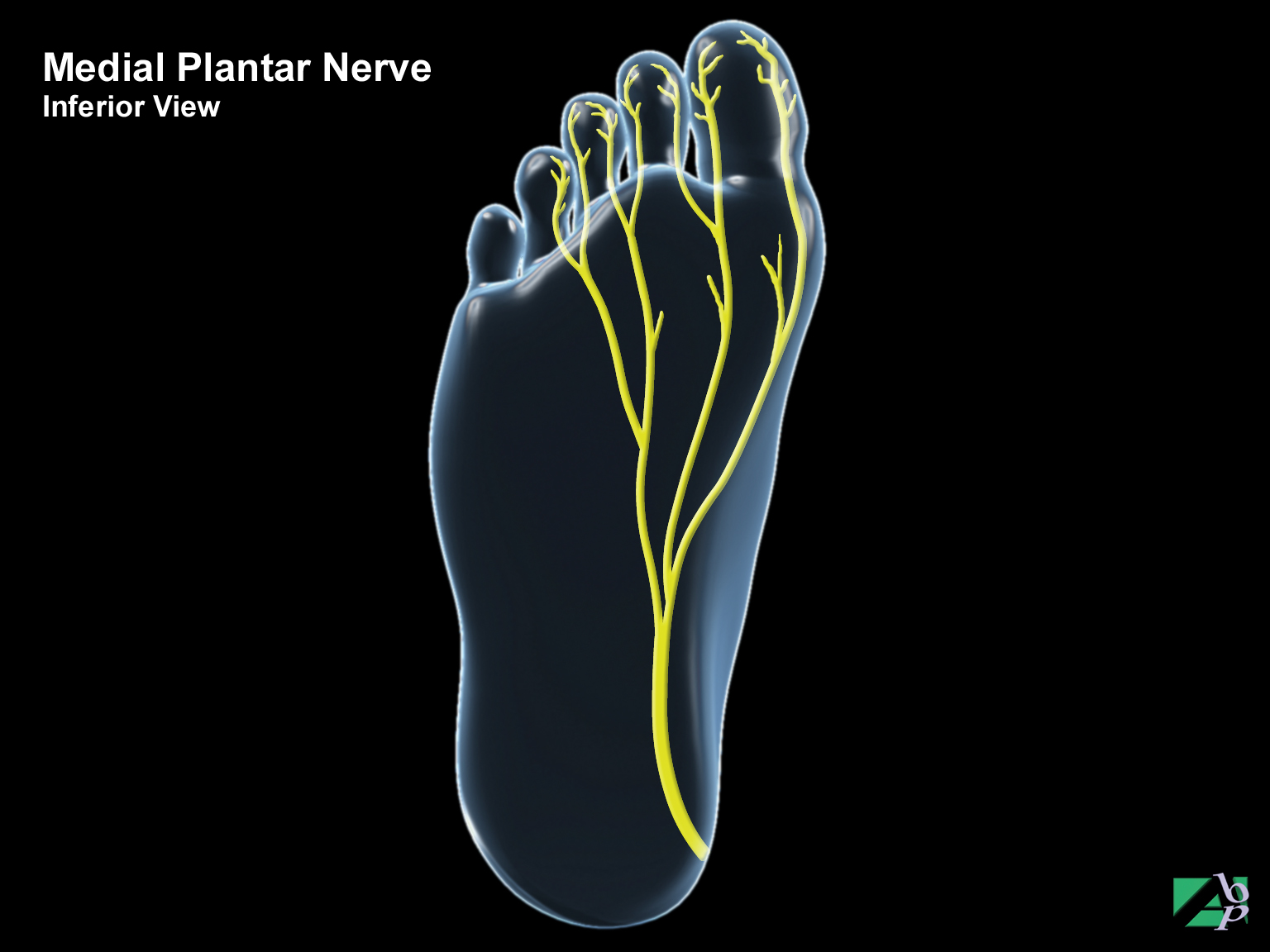
The medial plantar nerve is a terminal branch of the tibial nerve. It is a mixed nerve that supplies motor and sensory impulses to the foot and toes
Medial Rectus
An eye muscle originating from within the orbit and inserting onto the sclera. It adducts the eye. It is innervated by the oculomotor nerve
Median Nerve
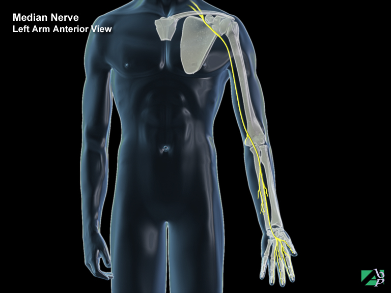
The median nerve is a mixed nerve (motor and sensory) that supplies motor branches to all the deep muscles of the forearm that flex the fingers and hand. A sensory branch originates just above the wrist, which supplies the skin of the palm and the base of the thumb. The main trunk of the nerve passes under the transverse carpal ligament to enter the palm. It supplies the intrinsic muscles of the hand and the thenar muscles of the thumb and supplies sensory branches to the sides of the thumb and the index, middle, and ring fingers, as well as the tips of these fingers and the soft tissues beneath the fingernails
Mediastinal
Relating to the mediastinum, the area between the lungs
Mediastinal Emphysema
Mediastinal emphysema is an abnormal condition in which there is air in the mediastinum (the space between the two lungs). It may be caused by a penetrating injury to the lung
Mediastinal Pneumogram
A graphical tracing of the movements of the chest showing the frequency, duration and depth of the respiratory movement
Mediastinum
The space between the lungs containing the heart, esophagus, trachea and vital blood vessels e.g., aorta, pulmonary artery
Medulla Oblongata
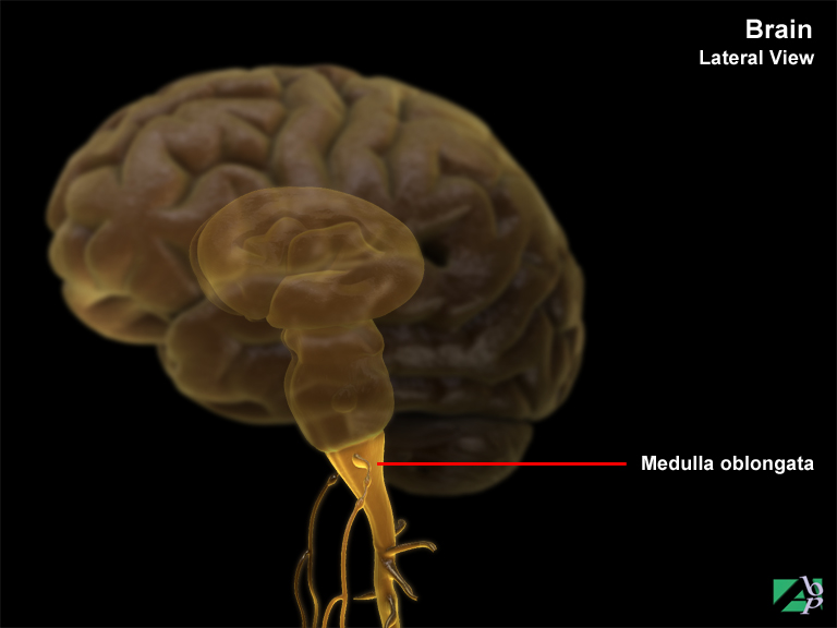
The medulla oblongata or as it is more often called, the medulla, is a bulbous structure that resembles the spinal cord and is continuous with the pons above and the spinal cord below. The medulla comprises vital nuclei and white matter that form the descending and ascending communication tracts between the spinal cord and the brain. Various cranial nerves arise from the medulla including the glossopharyngeal nerve, accessory nerve, hypoglossal nerve and vagus nerve. Other nuclei within the medulla function as autonomic centers for controlling vital visceral functions including heartbeat, arterial blood pressure and rate and depth of breathing
Medullary Canal
The canal that runs through the middle of a bone and contains the bone marrow
Meniere's Disease
A disease of the inner ear, the symptoms include vertigo, tinnitus and loss of hearing
Meningeal Adhesions
Meningeal adhesions are scar tissue on the spinal meninges, which are the membranes that surround and protect the spinal cord. The adhesions may form within the meninges on the arachnoid layer or on the outer layer, the dura. Trauma or surgery for trauma may be responsible for the adhesions either through inflammation of the meninges or through direct injury. Meningeal adhesions are likely to produce neurological symptoms in the arms or legs depending on the level of the spinal meninges involved
Meninges
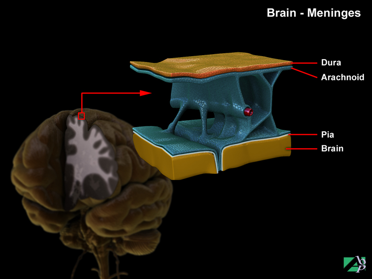
The term refers to the membrane that lines the spinal cord and the brain; there are three layers, the dura mater, the arachnoid and the pia mater. The pia is the innermost layer, the arachnoid the middle layer and the dura the outer layer
Meningitis
Inflammation of the meninges, which are the layers of membrane that line the spinal cord and the brain
Meniscectomy
This procedure is the removal of the meniscus from the knee. There are two menisci, a medial and a lateral meniscus. The medial meniscus is a crescent shaped cartilage that is found on the inner upper surface of the tibia. The lateral meniscus is another crescent shaped cartilage that is found on the outer upper surface of the tibia. Both cartilages form part of the knee joint
Mental Foramen
An opening in the mandible through which the mental nerve and blood vessels pass
Mentalis
A facial muscle in the chin area, it functions to elevate and protrude the lower lip
Meralgia Paresthetica
Neuralgic pain in the lateral aspect of the lower thigh, marked by symptoms of tingling and burning pain and some loss of sensation. It is caused by damage to the lateral femoral cutaneous nerve in the thigh
Mesencephalon
The midbrain, the middle portion of the brain
Mesenteric Artery
The mesenteric artery is a branch of the abdominal aorta, which supplies blood to part of the small intestine and part of the colon
Mesentery
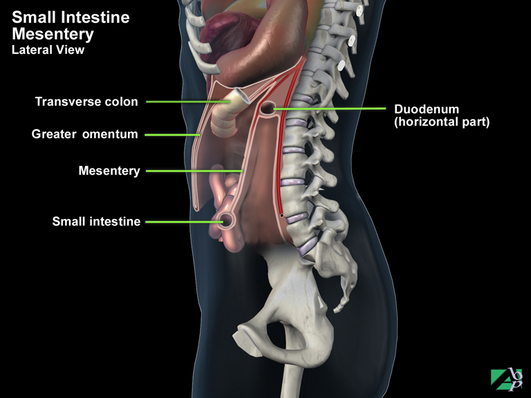
The mesentery is a double-layered peritoneal fold that connects the small intestine to the posterior wall of the abdomen. It supplies the nerve and blood supply to the small intestine and also allows it to alter shape and size
Mesocolon
A double layered peritoneal fold that connects the colon the abdominal wall
Metabolize
To convert
Metacarpal
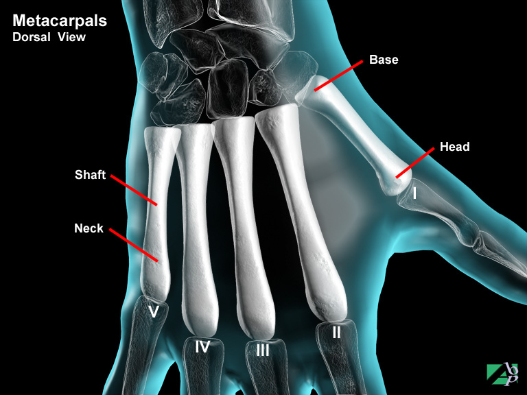
A bone of the hand, there are five in all. The metacarpal bones extend from the wrist (carpus) to the fingers. They are numbered one through five starting with the thumb side being the 1st metacarpal. A metacarpal bone has a head (the knuckle), a neck (just below the head), a shaft and a base. The metacarpal head articulates with a corresponding phalange (finger), while the metatarsal base articulates with a carpal bone of the wrist
Metacarpophalangeal
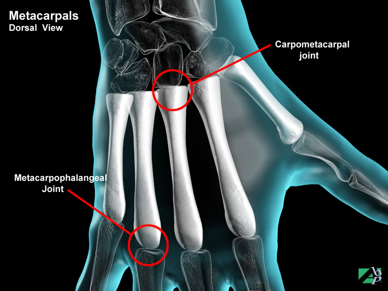
Refers to the joints between the metacarpal bones and the fingers (phalanges)
Metacarpus
The region of the hand where the wrist bones (carpus) articulate with the metacarpals
Metatarsal
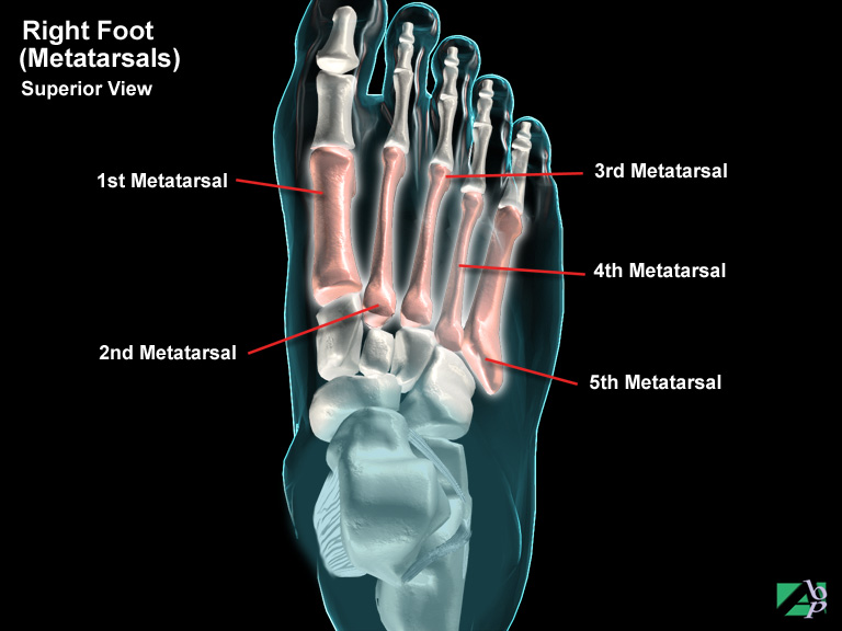
A bone of the foot, there are five in all. The metatarsal bones extend from the mid-foot (from the tarsal bones) to the toes (phalanges). They are numbered one through five starting with the big toe side being the 1st metatarsal. A metatarsal bone comprises a head, a shaft and a base. The metatarsal head articulates with a corresponding toe, while the metatarsal base articulates with a tarsal bone
Metatarsus
Referring to the mid-foot, that part of the foot between the tarsal bones and the toes, also known as metatarsus
Metatarsalgia
Pain in the mid part of the foot, the metatarsal area
Metatarsophalangeal
Refers to the joints between the metatarsal bones and the toes
Middle Ear
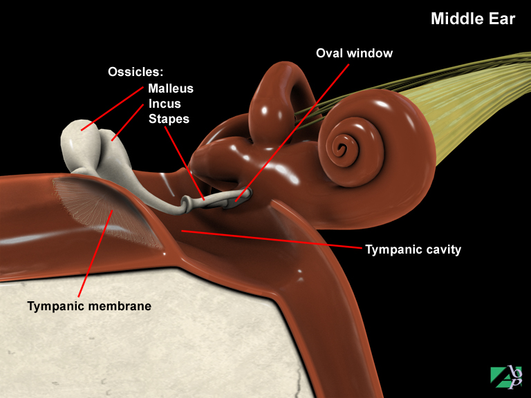
The middle ear is an air filled chamber called the tympanic cavity. The tympanic membrane (eardrum) separates the middle ear from the external auditory meatus. A bony partition containing what is referred to as the oval window and round window separates the middle ear from the inner ear. This bony partition is comprised of three small bones referred to as the ossicles. Individually these bones are named the malleus, incus and the stapes. These bones serve to amplify and transmit vibrations from the eardrum to the inner ear
Mid Foot
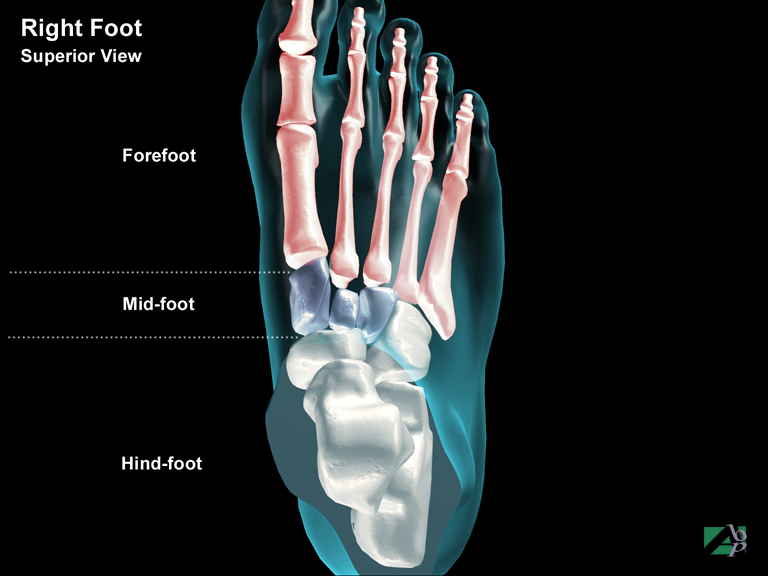
The middle part of the foot formed by the cuboid, navicular, and 1st, 2nd and 3rd cuneiform bones
Milwaukee Brace
An orthopaedic brace used to straighten the spine. It is used to treat scoliosis (abnormal side-to-side curvature of the spine)
Minerva Jacket
A body cast made of plaster of Paris, extending from just below the chin to the pelvis. It is used in the treatment of spinal fractures or dislocations
Monocular
Referring to one eye
Monteggia Fracture
A fracture dislocation of the forearm involving a fracture of the proximal ulna (near the elbow) and a dislocation of the radial head
Morbid Obesity
Extreme obesity
Mortice
A socket, the ankle mortice. A socket formed by the lateral and medial malleolus, which the talus fits into
Morton's Neuralgia
Morton's neuralgia or Morton's metatarsalgia is a painful condition caused by irritation of the plantar nerve, a nerve in the foot. It is associated with a neuroma, a growth on the nerve. The cause of the neuroma isn't clear but it is thought to be due to pinching of the nerve by the metatarsal heads when walking or standing. Ill-fitting shoes are often blamed for the condition
Motor Ataxia
Uncoordinated muscle movements
Mucobuccal Fold
The groove on the inside of the mouth between the gum and the chin
Muscles
The skeletal muscular system of the human body comprises over 600 individual skeletal muscles and in terms of body bulk makes up approximately 40% of the body's weight. Muscles essentially serve three purposes: body motion, body heat and body posture and support. There are three types of muscles within the body, smooth muscles, cardiac muscles and skeletal muscles. Skeletal muscles provide movement of the human body in response to stimulation from the peripheral nervous system; movement is achieved by contraction of the muscles in response to nerve stimuli
Muscles that attach to bone are said to have a point of origin and a point of insertion. Both ends of a muscle are attached to the bone by a tendon. A tendon is a tough fibrous tissue band that connects to the periosteum of the bone, which is the outer layer of the bone. Muscles generally function as units; muscles that contract together are referred to as synergistic. Antagonistic muscles perform opposite functions and are generally located on the opposite side of the limb
Musculoskeletal
Relating to the skeletal bones and the muscles that move the bones
Myelogram
A myelogram is a diagnostic spinal x-ray designed to evaluate any decrease or blockage in the flow of cerebrospinal fluid through the spinal column. It is used to diagnose spinal problems such as a bulging disc, tumor and bony abnormalities. The procedure is performed by first injecting a local anesthetic into the region of the spine to be x-rayed. A lumbar puncture or spinal tap is then performed, followed by injecting a dye or contrast agent. X-rays are then taken
Myocardial Contusion
Myocardial contusion or heart contusion is bruising of the heart muscles. It usually occurs from severe blunt trauma to the chest causing the sternum to compress the heart against the spinal column. This trauma leads to an alteration in the heart cells fluid composition, which in turn lead to an alteration in the heart's electrical activity and leads to abnormal heart rhythm. This rhythm activity is usually temporary
Myofascial
Relating to fascia. Fascia is a tough membrane underneath the skin covering the muscles
Myopia
Near sightedness
Myositis Ossificans
A condition in which bony substances deposit themselves within muscles
Myotomy
The surgical incision into a muscle
Myringitis
Inflammation of the eardrum
Myringoplasty
Reconstructive (plastic) surgery on the eardrum