Medical Glossary - Letter E
This medical glossary of terms beginning with the letter "E" contains the more common medical terms one might expect to encounter in a medical report or in hospital notes. The glossary is intended as a quick reference only; many of the terms are also referenced and illustrated in more detail in the medical libraries, to which you should refer for more detailed information.
Eardrum
The tympanic membrane, the membrane that separates the outer ear from the middle ear
Ecchymosis
Tiny hemorrhages under the skin, usually caused by trauma to the capillary blood vessels
Ectropion
Describes a condition where the edge of an eyelid is turned out
Eczema
A skin disease manifested by inflammation of the skin and watery blisters and itching
Edema
Swelling of tissue due to an accumulation of fluid
Edentulous
Without teeth, same as edentate
Efferent Nerve Impulses
Efferent nerve impulses are impulses generated by neurons from the Cental Nervous System and which are transmitted from the brain or spinal cord to a structure of the body such as a muscle or gland
Effusion
A discharge of fluid from tissue
Elbow
Three bones, the distal humerus and proximal ulna and proximal radius form the elbow joint. It is a hinge joint that allows rotational movements of the forearm. The elbow actually is comprised of three joints: the humeroulnar joint, in which the distal end of the humerus and the proximal end of the ulna connect; the humeroradial joint, in which the lower end of the humerus and the upper end of the radius connect; and the radioulnar joint, in which the upper ends of both the radius and the ulna connect. These joints are enclosed in what is referred to as a joint capsule, which provides support for the joint. The joint capsule is a membranous structure that completely encloses the articulating surfaces between the ulna and the humerus and the radial head and the humerus. A synovial membrane, which secretes synovial fluid, lines the capsule on its inner side to keep it lubricated. Joint support is also provided by a series of ligaments. These are the ulnar collateral ligament, radial collateral ligament and the orbicular ligament. In addition to these ligaments, strong muscles and tendons also help stabilize the joint and mediate the movements of the elbow and forearm
Electrocardiogram
A tracing of electric currents generated by the heart muscles, usually abbreviated to just ECG
Electroencephalogram
A tracing of electric currents generated by the brain. Also known as an EEG
Electromyography
Electromyography is the study and measurement of electrical activity of muscles. The muscle is electrically stimulated and a machine called an electromyograph measures the activity generated. The measurement is taken after electrodes are placed on the muscle
Embolectomy
The surgical removal of an embolism
Embolism
The blocking of a blood vessel. The blockage may be due to a blood clot, air or fat. The clot is called an embolus
Eminence
A projection on the surface of a bone
Empyema
A collection of pus in an organ or body cavity
Endarterectomy
An endarterectomy is the surgical removal of the intima, which is the innermost layer of an artery. It is usually performed to remove a thrombus (clot)
Endarteritis
Inflammation of the inner layer of an artery
Endocrine System
The glandular system of the body, which includes the pituitary gland, thyroid gland, adrenal glands and pancreas
The pituitary gland is a small structure about the size of a pea. Its main function is the production of a number of hormones, among these being growth hormones that stimulate growth of the body and which also influence the metabolism of carbohydrates, fats and proteins. It also produces hormones that control the function of the thyroid and adrenal glands and hormones that stimulate the gonads, ovaries and testicles. In addition, it stimulates the production of milk. Other hormones produced affect the contraction of the smaller blood vessels and the output of urine
The pancreas is located below the stomach between the liver and the spleen and behind a portion of the large intestine, roughly at the level of the twelfth thoracic vertebra. It is approximately 12.5 centimeters long and 2.5 centimeters thick. It is comprised of four sections: a head, neck, body and tail. Its function is vital to the metabolic processes of the body. Its secretions are essential to intestinal digestion and to insulin production that enables the body to absorb and metabolize (transform) glucose. The pancreas has two separate functional systems known as the pancreatic exocrine system and the pancreatic endocrine system. The pancreatic exocrine system produces three enzymes that pass through a duct system and which are secreted into the duodenum. This pancreatic juice contains water, bicarbonate and a variety of digestive enzymes that are used by the duodenum to digest food passed through by the stomach. About 98% of the pancreas's cell tissues are engaged in production of these digestive enzymes. The pancreatic endocrine system functions to produce insulin and other hormones. The insulin producing cells are located in an area known as the islets of Langerhans. Insulin regulates the body's uptake of glucose
There are two adrenal glands, which are situated just above each kidney. Each gland is composed of an internal portion called the medulla and an external portion called the cortex. The medulla produces a hormone called epinephrine, which raises blood pressure. It also stimulates heart rate, dilates coronary blood vessels and increases the respiratory rate. The cortex produces a group of very important steroid hormones including corticosterone, which influences the body's utilization of sugar, desoxycorticosterone, which controls the retention of sodium and water and aldosterone, which affects the utilization of sodium and potassium. In addition the cortex produces sex and reproduction hormones. The adrenal glands also produce hormones that assist the body to react to stress. The adrenal glands are stimulated by the sympathetic nervous system
The thyroid gland is located in the neck. It consists of two lobes connected by a narrow segment called the isthmus. The lobes lie on either side of the trachea (the windpipe). It secretes hormones that are necessary for growth and proper body metabolism. The major hormone produced by the thyroid is thyroxine, which is reliant on a sufficient intake of iodine and on thyroid stimulation by a hormone produced by the pituitary gland. Metabolic disorders occur when the rate of thyroxine production is too little or too much. Too little thyroid secretion due to insufficient intake of iodine results in hypothyroidism, too much secretion results in a condition known as hyperthyroidism resulting in weight loss, rapid pulse and protrusion of the eyeballs and an enlargement of the gland itself
Endolymphatic Shunt
This procedure is performed in order to drain excess fluid from the inner ear. A tube is inserted through the mastoid portion of the temporal bone into the endolymphatic sac, a duct in the inner ear. It is usually performed to give relief to vertigo
Endoscope
A tubular instrument used for the visualization of the interior of an organ, it usually has an optical lens and light at one end
Enema
An enema is an injection of fluid through the rectum into the large intestine. It is normally used to facilitate emptying the bowel in cases of constipation but it may also be a way of providing nutrients and is also used as an aid in diagnosing ailments of the large intestine
Entropion
An entropion is an abnormal infolding of the edge of the eyelid with the result the eyelashes rest against the surface of the eye
Epicondyle
A prominence on a long bone (eg. the humerus) situated above a condyle, the latter being an expansion at the end of a bone for articulation with another bone
Epicondylitis
Inflammation of an epicondyle and / or its tendinous attachments. An epicondyle is a bony projection on the lower end of some long bones such as the femur and humerus. The epicondyle is the attachment point for muscle tendons
Epidermis
The outer layer of the skin, it is without nerve endings or blood vessels, although sweat glands and hair follicles pass through it. The epidermis consists of five separate layers of cells. All but the deepest layers are actually dead cells that contain a protein, which toughens and waterproofs the skin. These cells form in the bottom layer of the epidermis and are pushed outward to the surface where after a period of time they are transformed into hard flakes, which form a dry outer coating of the skin. This crust flakes off and is continually being replaced with new merging cells. The epidermis is not subject to scarring from injury or exposure
Epidural Hematoma
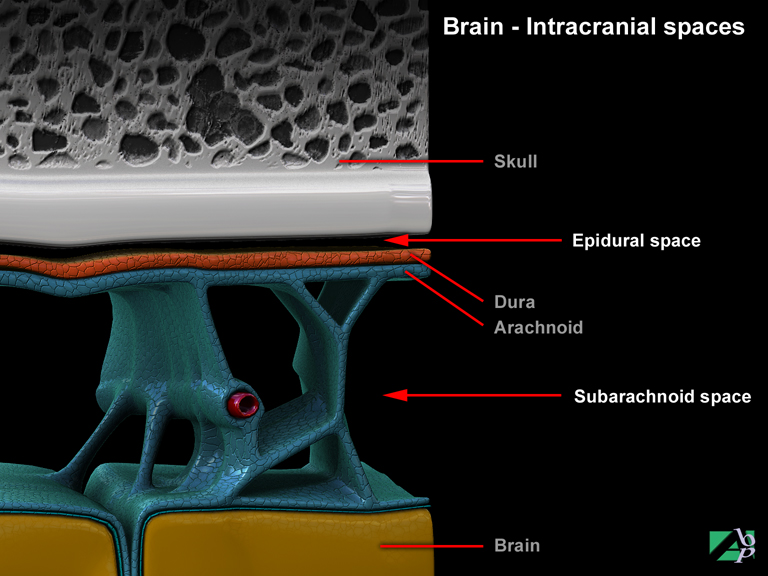
Epidural hematoma or as it is sometimes called extradural hematoma, develops between the outer dural layer and the inner table of the skull in the potential space called the epidural space. Normally there is no epidural space as the dura mata is attached to the inner skull but a space can be created if the dura is stripped away from the skull such as might be seen following a fracture. Hemorrhage of blood vessels cause blood to accumulate in the epidural space forming a hematoma. As the hematoma expands it causes intracerebral pressure that compresses the brain. Unless this pressure is relieved, brainstem herniation occurs crushing the midbrain and resulting in death
Epidural Space
The epidural space is a space or potential space between the inside of the skull and the dura mater, the outer layer of the meningeal lining covering the brain and the spinal cord
Epigastric Region of Abdomen
The epigastric region of the abdomen is the upper median portion of the abdomen. It contains portions of the liver, stomach, pancreas and the duodenum
Epiglottis
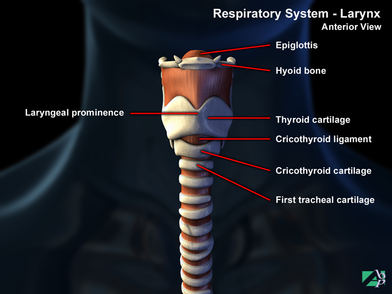
A piece of tissue that serves as a cover for the larynx during the act of swallowing
Epilepsy
Epilepsy may be best simply described as a sudden temporary disturbance of brain function sometimes accompanied by altered states of consciousness and violent seizures or convulsions. It is caused by a malfunction of the electrical activity of the neurons of the brain. The cause of epilepsy is not clear but there is a clear nexus between epilepsy and head trauma
There are various stages of epilepsy. An early stage is known as an aura although this is not always experienced in an epileptic episode. An aura describes sensory symptoms such as ringing in the ears or seeing spots before the eyes. Following the aura is the seizure that is known as the ictus, a.k.a. a convulsion. The convulsion stage varies depending on the type of epileptic seizure suffered. Convulsion signs may vary from loss of consciousness to a staring spell. The time between the epileptic episode and return to normal consciousness is referred to as the postictal stage. Epileptic seizures fall into various categories that are described according to their symptoms and frequency; however the most common form is referred to as generalized seizures
Generalized seizures result from abnormal electrical disorders affecting the entire cerebrum. There are numerous forms of generalized epilepsy but this type includes two of the most common posttraumatic forms: petit mal and grand mal epilepsy. Petit mal epilepsy is the most common form of generalized epilepsy seen in children. It is rare in adults. Seizures can occur infrequently or as many times as several hundred per day. In this form there is no loss of consciousness, only a brief alteration in consciousness. Grand Mal epilepsy is the most common type of generalized seizure. This is sometimes referred to as tonic/clonic type. In this type of seizure a profound and abrupt loss of consciousness occurs along with contractions of large muscle groups that extend the arms and legs and arch the back, resulting in violent spasms or jerks of the whole body
Epiphyseal Fracture
A fracture of the end of the shaft of a bone where the epiphyseal plate is located, the epiphyseal plate is growth area of a long bone. This type of fracture only affects children or adolescents, in adults the epiphyseal plate closes and the cartilage is replaced by normal bone
Epiphysis
The epiphysis is a section of cartilage at the end of the shaft of a long bone that is seen during childhood and adolescence. This cartilage is where the growth of the long bone takes place. Later in life when growth has ceased, the epiphysial cartilage is displaced by a bony fusion of the head of the long bone and the shaft
Epistaxis
Nosebleed
Epithalamus
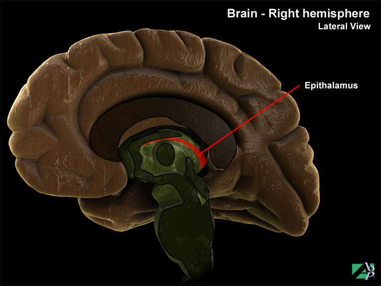
The epithalamus is the dorsal (back) portion of the diencephalon (a small part of the brain situated between and below the cerebral hemispheres and above the medulla oblongata and the cerebellum). The roof of the epithalamus consists of a vascular choroid plexus where cerebrospinal fluid is produced
Epithelium
The epithelium is a thin layer of tissue that covers all surfaces of the body, internal organs, blood vessels, glands etc
Equine Gait
A term used to describe an abnormal gait, a high stepping gait, in which the foot is raised higher off the ground than is normal or necessary. It may be due to a peripheral nerve lesion involving the peroneal nerve
Erb's Palsy
Partial paralysis of the muscles of the upper arm due to injury to the brachial plexus
Erector Spinae Iliocostalis
A spinal muscle originating from the ribs and inserting onto the transverse processes of the vertebrae above and below its point of origin. It assists extension and lateral flexion of the spine
Erector Spinae Longissimus
A spinal nerve running along much of the length of the spinal column. It assists extension of the spine
Erector Spinae Spinalis
A spinal muscle, it assists lateral flexion of the spine
Esophagus
The esophagus (the gullet) is part of the gastrointestinal track. It is a tubelike structure that forms a canal between the mouth and the stomach. Its upper end connects with the pharynx. It descends behind the trachea and passes through the thoracic diaphragm (the mediastinum) and then through an opening called the esophageal hiatus into the stomach. It is formed primarily of muscle. Its function is to transport food to the stomach. It plays no role in the breakdown or absorption of food
Ethmoid Bone
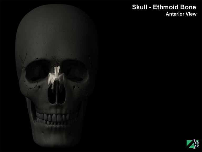
One of the bones of the skull, it is located in the anterior portion of the floor of the cranium between the orbits where it forms the roof of the nasal cavity
Etiology
The cause of a disease or disorder
Eustachian Tube
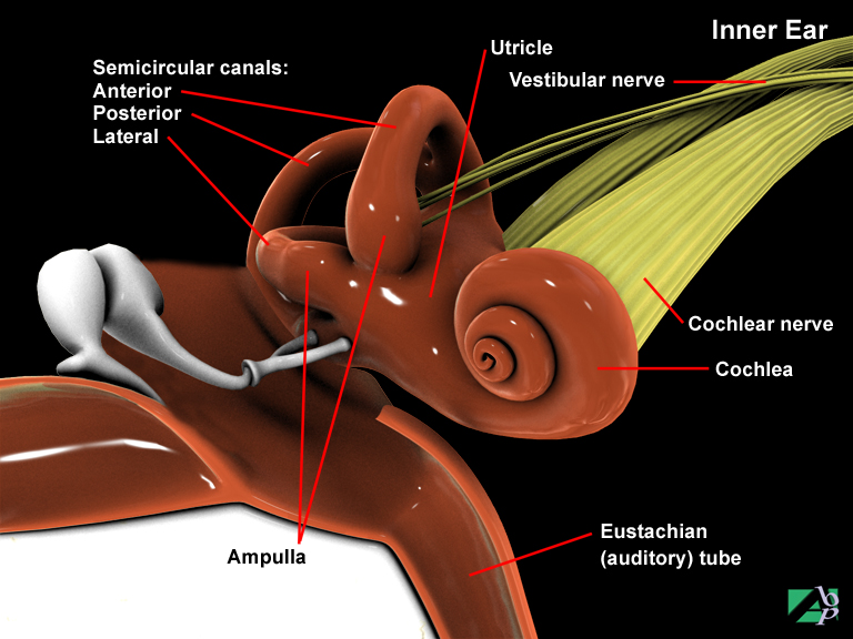
A canal that communicates between the chamber of the middle ear and the pharynx. Its purpose is to equalize the pressure in the middle ear to that of the atmosphere outside
Eversion
Turning outward, it is a term usually applied to a deformity of the foot, or to describe movement of the foot at the ankle joint, or the outward turning of an eye
Evisceration
This term applies to the surgical removal of an eyeball
Extension
Extension is a term used to describe movement of part of a limb, for example to release a grasped object it requires extending or straightening the fingers to release the object. Extension is the opposite movement to flexion. In the example given, to grasp an object it requires flexion or bending the fingers to grip the object. Muscles that allow extension of a body part are known as extensor muscles, and the tendinous attachments as flexor tendons
Extensor Carpi Radialis Brevis
A muscle running along the inside of the forearm. It originates from the lateral epicondyle of the humerus at the elbow and inserts onto the base of the 3rd metacarpal of the hand. It extends and abducts the hand at the wrist
Extensor Carpi Radialis Longus
A muscle extending along the inside (thumb side) of the forearm. It originates from the humerus at the elbow and inserts onto the base of the 2nd metacarpal of the hand. It extends and abducts the hand at the wrist. The radial nerve innervates it
Extensor Carpi Ulnaris
A muscle extending along the outside (little finger side) of the forearm. It originates from the lateral epicondyle of the humerus just above the elbow and inserts onto the 5th metacarpal of the hand. It extends and adducts the hand at the wrist
Extensor Digiti Minimi of Hand
A muscle extending from the lateral epicondyle of the humerus, which inserts onto the little finger of the hand. It provides for extension of all of the joints of the little finger
Extensor Digiti Minimi of Foot
A muscle of the foot originating from the heel (calcaneus) and inserting onto the big toe, and the 2nd, 3rd and 4th toes. It extends the toes when the foot is dorsiflexed
Extensor Digitorum of Hand
A muscle originating from the humerus near the elbow region extending down the forearm to the fingers. It assists extension of the fingers
Extensor Digitorum Longus of Foot
A muscle extending down the lower leg, originating from the fibula and attaching to the 2nd, 3rd, 4th and 5th toes. It extends the toes and the foot at the ankle. The deep peroneal nerve innervates it
Extensor Hallucis Longus
A muscle extending from the mid shaft of the fibula (from the front of the middle part of the lower leg) extending down the leg into the foot and inserting onto the big toe. It helps extend the big toe and the foot and assists inversion of the foot. The deep peroneal nerve innervates it
Extensor Indicis
A muscle originating from the shaft of the ulna in the forearm and extending into the hand and inserting onto the index finger. It provides extension of all of the joints of the index finger
Extensor Pollicis Brevis
A muscle extending from the shaft of the radius down into the hand and inserting onto the base of the thumb. It assists extension of the metacarpophalangeal joint of the thumb
Extensor Pollicis Longus
A muscle of the forearm originating from the upper end of the ulnar extending down the forearm and inserting onto the distal phalanx of the thumb. It extends the interphalangeal and metacarpophalangeal joints of the thumb
External Auditory Canal
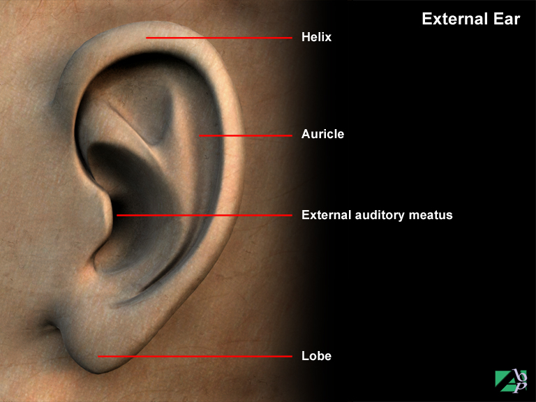
The canal or opening in the external ear
External Carotid Artery
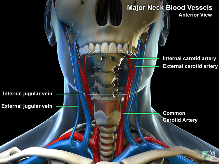
The external carotid artery is a branch of the common carotid artery. It supplies blood to the head
External Fixation
External fixation refers to the stabilization of a fractured bone after it has been reduced. Devices sometimes referred to as external fixators are used to hold (immobilize) reduced fractures. Externally placed plates or rods are used to keep the reduced fracture in anatomical alignment. The plates or rods are held in place by screws or pins inserted into the bone above and below the fracture site
External Jugular Vein
The external jugular vein is one of two veins on each side of the neck: the other is the internal jugular vein. The external jugular is the smaller of the two veins. It drains blood from parts of the face and the scalp into the subclavian vein
Extraluminal
This term refers to the outside of a hollow structure or organ, i.e., the esophagus or an artery. The term may be used to describe an injury for example an extraluminal injury to the esophagus, which would mean it was an injury to the outer surface of the esophagus, such as from a laceration
Exudation
Oozing of fluid from the body
Eye
The eye consists of three layers or tunics. These are known as the fibrous layer, the vascular layer and the internal or neural layer. The outer fibrous layer is divided into two regions comprising the opaque sclera and the cornea, which is transparent. The vascular layer is known as the uvea. It comprises the choroid, the ciliary body and the iris and lens. The internal layer contains the retina
Eyelid
The eyelids are vital to the eye's structural defences. Their natural pattern of blinking every few seconds creates a strong barrier between the eye and the external environment. Their reflexive closure in response to threat to the eye also acts as a barrier against injury to the eye. The act of blinking also lubricates the eye by spreading tears formulated in glands in the eyelids across the surface of the eye. The inner surface of the eyelid is lined with a thin mucous membrane called the conjunctiva; this allows the eyelid to pass over the eye without friction. The eyelid also has a row of numerous eyelashes that protect the eye from airborne objects. Eyelids of the upper lid are long and turn upwards whereas those of the lower lid are short and turn downwards