Medical Glossary - Letter S
This medical glossary of terms beginning with the letter "S" contains the more common medical terms one might expect to encounter in a medical report or in hospital notes. The glossary is intended as a quick reference only; many of the terms are also referenced and illustrated in more detail in the medical libraries, to which you should refer for more detailed information.
Sacral
Refers to the sacral area, the lower back
Sacral Plexus
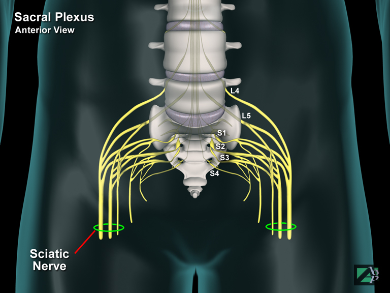
The sacral plexus is a network of nerves formed by the spinal nerves that arise from L5, S1, S3 and S4. A major peripheral nerve arising from this plexus is the sciatic nerve, which innervates the skin and muscles of the leg and foot
Sacrococcygeal
Relating to the sacrum and coccyx region
Sacrococcygeal Ligament
A ligament that binds the sacrum and coccyx together
Sacroiliac
Relating to the sacrum and ilium, the upper part of the pelvis
Sacroiliac Joint
The sacroiliac joint is the articulation between the sacrum and the ilium of the pelvis
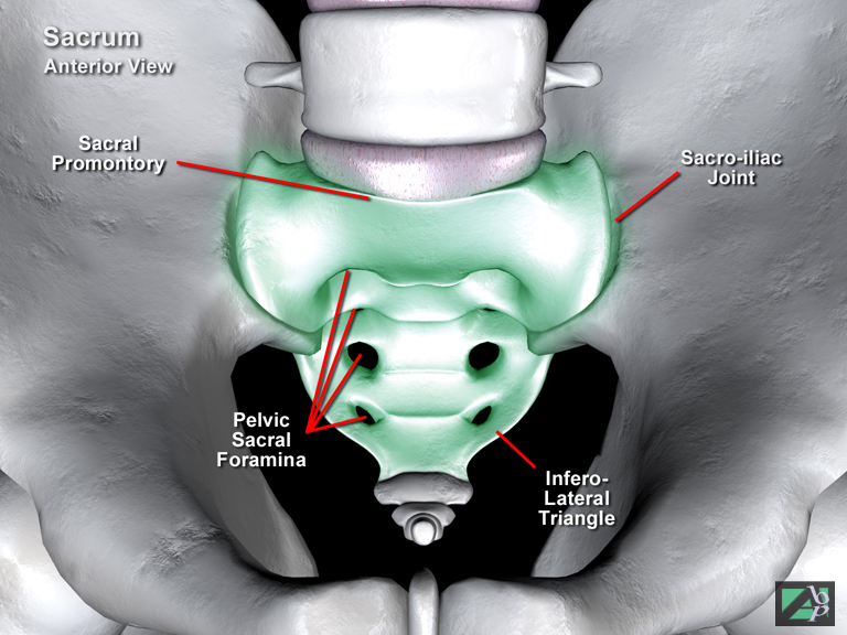
Sacrum
The sacrum is a wedged shaped bone, which begins as five separate vertebrae, which fuse around the age of 25 years. It provides strong support for the pelvic girdle and actually forms part of the pelvis. It fits into a wedge shaped opening between the back ends of the two hip bones (the ilium) where it is known as the sacroiliac joint. The cauda equina of the spinal cord runs within the central canal of the sacrum and spinal nerve roots exit through foramina contained within the sacrum, notably the sciatic nerve exits from the sacrum
Sagittal Suture
The regions of the skull are divided by membranous and cartilaginous tissue called suture. The sagittal suture divides the frontal and parietal bones
Salmonella
Salmonella is form of bacterium. There are three forms of Salmonella infection that occur in humans: typhoid (enteric) fever, bacteremia (bacteria in the blood stream) and acute gastroenteritis, which is the most common form. Most cases of salmonella poisoning generally results from ingestion of contaminated food or water. Poultry and poultry products and beef are the main sources of salmonella poisoning. Salmonella primarily affects the gastrointestinal tract. Symptoms include nausea and vomiting, aching muscles, fever, headache and diarrhea
Saphenous Nerve
A branch of the femoral nerve. It provides sensation to the inner surface of the leg and the foot
Saphenous Veins
The great saphenous vein commences in the arch of the foot and ascends along the inside of the leg to the thigh where it drains into the femoral vein. The small saphenous vein commences from the outside of the foot and ascends the back of the leg to the knee where it drains into the popliteal vein
Sartorius
A muscle of the upper leg originating from the iliac spine of the pelvis and inserting onto the upper shaft of the tibia. It assists flexion, abduction and lateral rotation of the thigh at the hip and medial rotation of the leg at the knee. It is innervated by the femoral nerve
Scalenus Anterior
A muscle in the back of the neck originating from the transverse processes of C3-C6 and attaching to the 1st rib. It assists breathing and lateral flexion of the neck
Scaphoid
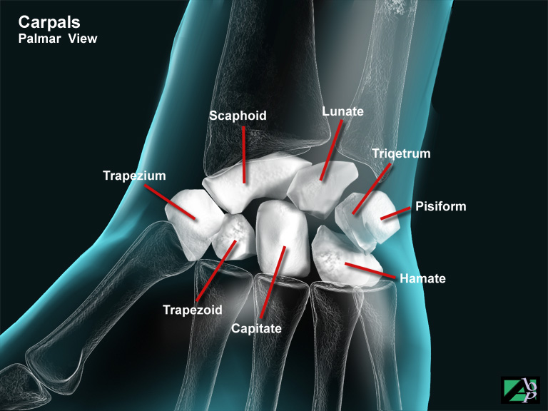
A carpal bone, i.e., a bone of the wrist and in terms of wrist function is the most important of the carpal bones. It connects with the styloid process of the radius and with the adjacent lunate bone and with the trapezium, trapezoid and capitate bones. The scaphoid is sometimes referred to as the navicular bone
Scapula
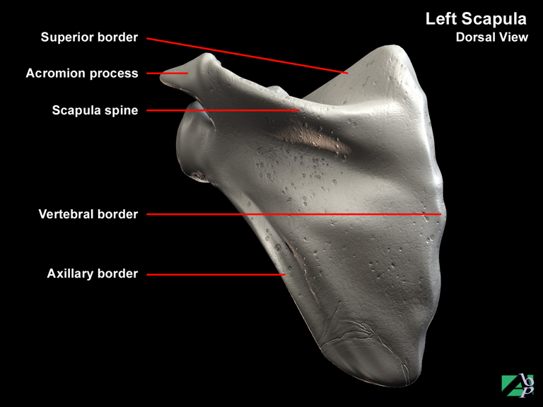
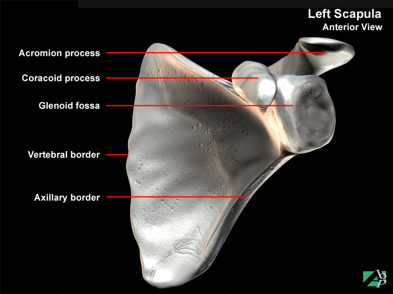
The scapula or shoulder blade is a triangular shaped bone in the back of the shoulder. The scapula has three borders separated by two angles. The upper border is called the superior border. The border nearest the vertebrae is called the medial or vertebral border and the border facing towards the arm is called the axillary border. It is also comprised of a spine which is a prominent bony ridge running diagonally across the bone. Towards the shoulder, the spine broadens and becomes the acromion process, which allows for movement with the clavicle. Below the acromion process is a depression called the glenoid cavity into which the head of the humerus fits. Near to this cavity is another bony process called the coracoid process, which gives attachment to several shoulder ligaments. In addition to these articulations the scapula is an important point for the insertion of muscles that assist motion of the shoulder
Schmorl's Nodes (Nodules)
Small lumps evident on spinal x-rays that are indicative of prior intervertebral disc herniation, they are formed by the herniated disc material, the nucleus pulposus
Sciatic Foramen
An opening in the sacrum through which the sciatic nerve emerges
Sciatic Nerve
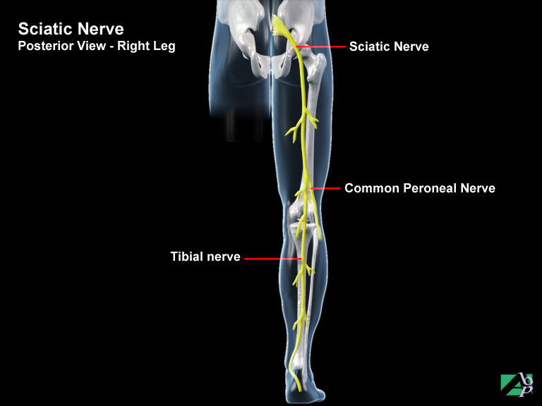
The sciatic nerve is a mixed nerve (motor and sensory) and is the largest peripheral nerve in the body. It passes from the pelvis through an opening called the sciatic foramen and emerges in the buttock area and extends down the back of the thigh to the knee where it divides into two nerves, the tibial nerve and the common peroneal nerve. In the thigh region the sciatic nerve innervates (moves) the muscles of the hamstrings (the biceps femoris, semitendinosus, semimembranosus muscle and the adductor magnus). These muscles extend the thigh and flex the knee joint. Total lesions of the sciatic nerve above the knee where it is one nerve will also affect the innervations and sensory supply of the tibial and common peroneal nerves
Sciatica
Sciatica is a somewhat vague term used to describe sensory disturbances in the leg and foot. The principal symptom is pain. The symptoms may be due to a sciatic nerve lesion, but more often than not, they are due to intervertebral disc disease
Sclera
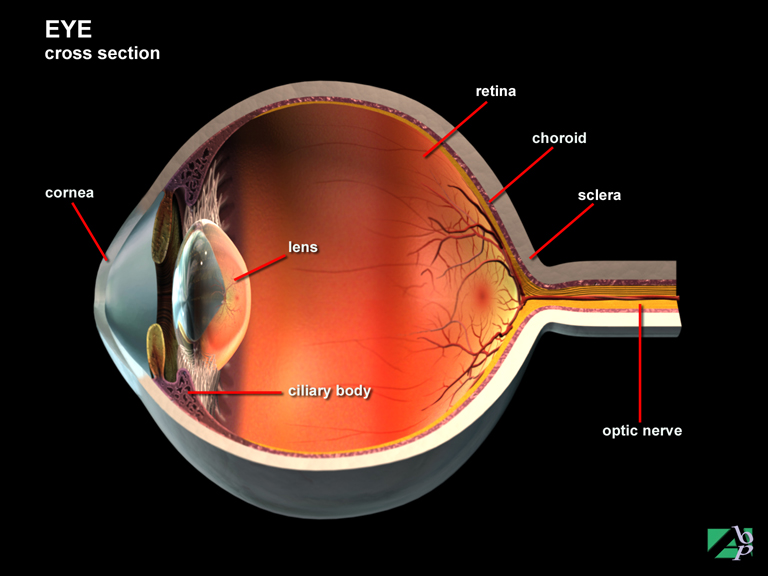
The sclera is the white part of the eye. It is made up of elastic and collagenous fibers that serve to give the eyeball its shape and to protect the inner structures of the eye
Scleral Buckling
Scleral buckling is a technique used for retinal detachment. A tight band is placed around the eyeball causing it to buckle, which brings the sclera and choroid inwards and in contact with the detached retina. When contact is made, diathermy, cyrothermy or photocoagulation is then applied to permanently fuse the retina and choroid together.
Scoliosis
An abnormal curvature of the spinal column, lateral or medial ie. to either side
Scrotum
The scrotum is a sac like structure suspended behind the base of the penis. It functions to support and protect the testes and to regulate their position. Muscles within the subcutaneous layers of the scrotum allow the scrotum to contract or expand according to environmental conditions (heat and cold), which position the testes so that an even temperature is maintained which is necessary for production and storage of sperm. The scrotum is also divided into two compartments by a fibrous septum. This septum, the median septum serves to compartmentalise the testes so that injury or infection will only affect one or the other, not both. The pudendal nerve, the ilioinguinal nerve and the posterior cutaneous nerve, all of which are primarily sensory nerves, innervate the scrotum
Second-degree-burn
Second-degree burns involve the entire epidermis and part of the dermis. Second-degree burns are sometimes described as superficial and deep 2nd degree burns. Superficial burns involve the entire epidermis and the outermost layers of the dermis. Deep burns extend deep into the dermis. Superficial 2nd degree burns are blisters and are red in appearance. After the initial burn the skin may appear to weep. Although these burns are painful they usually heal within a few weeks. Second-degree superficial burns do not result in scarring, however some minor pigmentation may occur and the area burned may appear a slightly different color to the surrounding unburnt skin. Deep 2nd degree burns are likely to result in scarring and are more prone to secondary infection
Sella Turcica
A depression in the sphenoid bone of the skull, which houses the pituitary gland
Semilunar Bone
Another name for the lunate bone, one of the carpal bones of the wrist
Semilunar Cartilage
The meniscus, either of the lateral or medical meniscus of the knee joint
Semimembranosus
A muscle of the upper leg originating from the ischial tuberosity of the pelvis and attaching to the medial condyle of the tibia. It assists flexion and medial rotation of the knee and extension of the hip. It is innervated by the tibial nerve
Semitendinosus
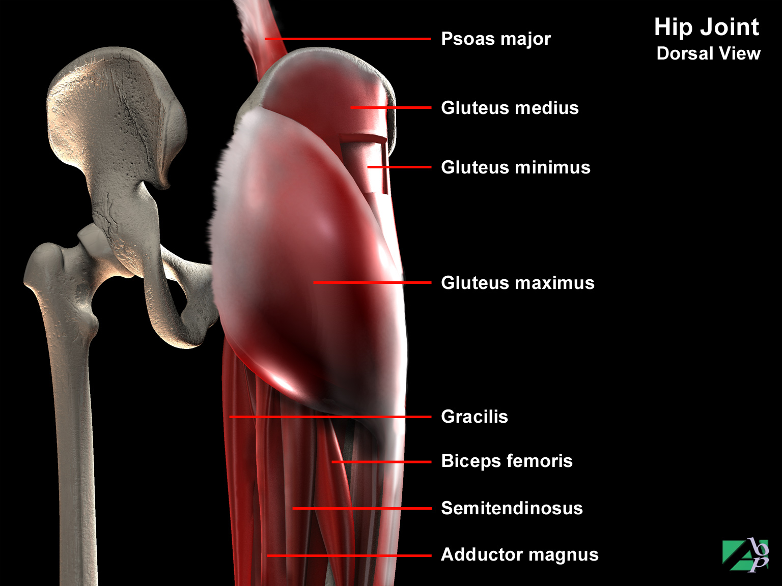
A muscle of the upper leg originating from the ischial tuberosity of the pelvis and inserting onto the upper shaft of the tibia. It assists flexion and medial rotation of the knee and extension of the hip. It is innervated by the tibial nerve
Sensory Nerve Impulses
Incoming neural impulses are called afferent impulses or sensory impulses. These sensory impulses are initiated at the peripheral receptor organs of the body: the skin, subcutaneous tissue, muscles, ligaments, internal organs and organs of the special senses. Afferent impulses occur in response to outside or environmental conditions and internal conditions within the body itself such as pain, heat, itching
Sepsis
An infection
Septoplasty
Septoplasty, or submucous resection is usually performed to correct a deformity of the nasal septum. It involves making a small incision inside the nose and detaching the mucosal lining from the bones and cartilage. The deformed part of the septum is then straightened or removed and the mucosal lining re-attached. Splints or packs are then placed in the nose
Sequestrectomy
Sequestrectomy is the surgical removal of a dead piece of bone known as a sequestrum
Sequestrum
A piece of dead bone
Serratus Anterior
A muscle of the upper back originating from the intercostals of the first eight ribs and attaching to the scapula. It assists lateral rotation and protraction of the scapula
Sesamoid Bone
A sesamoid bone is a bone that lies within a tendon that moves over a joint surface, the patella for example is a sesamoid bone. In the foot there are at least two sesamoid bones, the tibial and fibular sesamoid, which are located in the flexor hallucis brevis below the head of the first metatarsal. Some individuals may have more than these two sesamoid bones
Sesamoidectomy
Excision of a sesamoid bone in the foot, one of two bones that lie below the head of the 1st metatarsal bone
Shoulder
The articulation between the clavicle, scapula and the humerus
Shoulder Joint
The articulation by the head of the humerus into the glenoid cavity of the scapula
Sialoadenectomy
Sialoadenectomy is the surgical removal of a salivary gland. It may be total or partial. The major salivary glands are the parotis gland, sublingual gland and the submaxillary gland
Sigmoid Colon
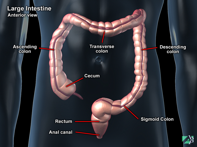
The last part of the colon, the last part of the large bowel
Sigmoidectomy
Sigmoidectomy is the surgical removal of part or all of the sigmoid, which is the terminal end of the colon
Silastic
Refers to silicone or elastic or rubber material
Sinogram
A sinogram is a contrast x-ray of a sinus, which refers to a large channel or an air space. Sinography is used to x-ray regions such as the maxillary sinus and the other sinus's of the facial bones and also of the skull, but it is also used to x-ray fistula's (abnormal openings or channels), particularly of the chest and abdominal wall
Sinusotomy
Sinusotomy is a surgical procedure in which an incision is made into the nasal sinus, usually to discharge infected fluid or material
Skeletal Traction
Skeletal traction is a form of traction where the pull is exerted directly upon the fractured bone by means of attaching weights to pins or wires inserted through the bone. Skeletal traction is usually reserved for long-term traction and may be referred to as continuous traction. Skeletal traction applied for shorter periods, or re-applied may be referred to as intermittent traction
Skin
The skin is actually considered an organ, as it comprises several kinds of tissues that are arranged in such a manner as to function together. The primary function of the skin is to act as an impermeable (resistant) barrier between the external environment and internal tissues while assisting to hold tissues and fluids in place within the body. It also permits bodily communication with the environment through its sensory nerve distribution. The skin functions to protect the body from disease and external injury, it helps regulate body temperature and fluid retention. It synthesises vitamin D absorption and acts as the body's primary sensory receptor to the external environment responding to heat, cold, vibration, touch, pressure and other stimuli. The skin varies in thickness from body region to region. It is thickest where there is most wear and tear, the soles of the foot for example. The skin is composed of three structural layers: the epidermis, dermis and hypodermis (some consider the hypodermis to be part of the dermis). The epidermis is the thinnest, outermost layer; it is without nerve endings or blood vessels, although sweat glands and hair follicles pass through it. The epidermis consists of five separate layers of cells. All but the deepest layers are actually dead cells that contain a protein, which toughens and waterproofs the skin. These cells form in the bottom layer of the epidermis and are pushed outward to the surface where after a period of time they are transformed into hard flakes, which form a dry outer coating of the skin. This crust flakes off and is continually being replaced with new merging cells. The epidermis is not subject to scarring from injury or exposure. The dermis or corium lies beneath the epidermis and consists of two layers: the papillary layer and the reticular layer. The outer layers join the underside of the epidermis. It consists of many capillary blood vessels, sweat glands, nerve endings and hair follicles. The deeper and thicker layer consists principally of thick interwoven bundles of elasticized fibers, which allows the skin to stretch
Skin Traction
Skin traction is a form of traction where weights are applied using tape, sponge rubber or canvas material and attaching the weights to the skin around the fracture site. There are four basic types of skin traction used: Buck's traction, Russell's traction, Dunlop's traction and Thomas' splint traction. Buck's traction and Russell's traction are used for lower extremity fractures while Dunlop's traction is used for upper extremities fractures. Thomas' splint traction is used primarily for lower limb fractures but is sometimes also used for arm fractures. Skin traction has limitations, as the amount of weight that can be used is limited to the tolerance of the skin
Skull
The skull (cranium) bones are made up of eight cranial and fourteen facial bones. The skull contains numerous cavities these being the cranial cavity (the largest of all), the nasal cavity, middle and inner ear chambers and the two bony orbits. The latter that houses the eyeballs is actually formed by facial and cranial bones. The bones of the skull are partitioned into two parts, the vault and the base. The vault, which is also known, as the calvarium, is the upper rounded dome like portion. It is made up of the frontal bone, two parietal bones, two temporal bones, an occipital bone, a sphenoid bone and an ethmoid bone. The base refers to the irregular portion located below the vault. The base of the skull is made up of the basal portion of the frontal bone, the ethmoid, the sphenoid and the basal portion of the occipital bone. The contour of the base of the skull is irregular and rough as opposed to the contour of the vault of the skull that is a smooth convex surface. The skull base is also perforated with passages for cranial nerves and blood vessels to pass through. The base of the skull also contains the foramen magnum, which is a large opening of the occipital bone that provides a passageway for the lower portion of the brainstem, as it becomes the upper portion of the spinal cord. The vault and base are separated by what is termed sutures of the skull. These sutures are made up of membranous tissue and cartilage. The four prominent skull sutures are the coronal suture, the sagittal suture, the lamboidal suture and the squamosal suture. The coronal suture unites the frontal bone with the two parietal bones. The sagittal suture unites the bones at the midline. The lamboidal suture separates the parietal and occipital bones. The squamosal suture is located between the parietal and temporal bones
Small Bowel Series
This is an x-ray of the small bowel (the duodenum, jejunum and ileum)
Small Intestine
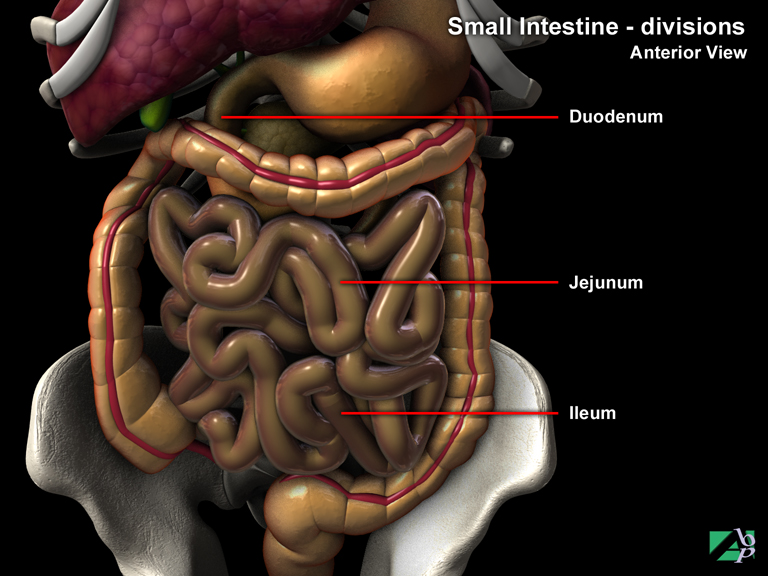
The small intestine is a coiled tube about 20 feet in length and is located in the center of the lower abdomen. The small intestine is divided into three arbitrary sections: the duodenum, the jejunum and the ileum, note however that these three sections are indistinct as the intestines are continuous. The duodenum begins at the pyloric opening with the stomach. It is C shaped and bound to the abdomen by the peritoneum (the lining of the abdomen). The duodenum is in close contact with several other abdominal organs including the liver, pancreas, gallbladder and left kidney. At about the level of the 2nd lumbar vertebra it becomes the jejunum. The jejunum segment is about nine feet long and is similar in appearance to the duodenum. The ileum is the last segment of the small intestine beginning at the jejunum and ending at the junction with the large intestine. It is smaller in diameter than the jejunum but longer and its walls are thinner and are less vascularized. Both the jejunum and ileum are completely covered by the peritoneum
Smith Peterson Nail
An internal fixation device, a nail used to secure proximal fractures of the femur ie. fractures of the femoral head or neck
Smith's Fracture
A Smith's fracture is a reverse Colles fracture; in this type of distal radial fracture the fracture results in palmar displacement
Snellen Test
A test for visual acuity
Soft Palate
The back part of the roof of the mouth, towards the throat
Soleus
A muscle of the lower leg originating from the tibia and fibula and inserting onto the calcaneus in the foot. It assists plantar flexion of the foot. It is innervated by the tibial nerve
Spasm
An involuntary muscle contraction
Sphenoid Bone
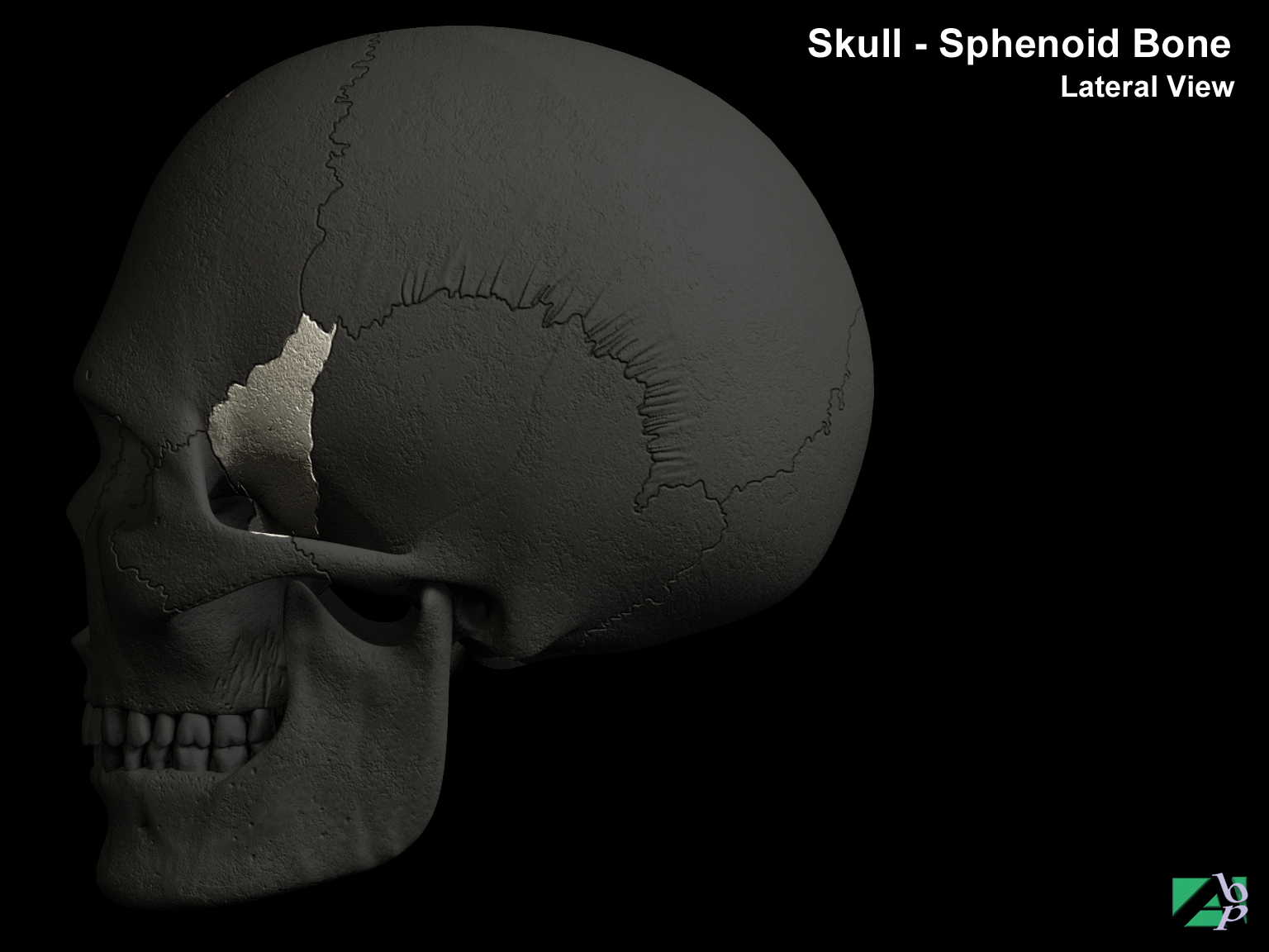
The sphenoid bone forms one third of the floor of the cranium. The ethmoid and frontal bones in front and the temporal and occipital bones at the back surround it. In the center of its inner surface is a depression that houses the pituitary gland
Sphenoid Electrodes
Sphenoid electrodes are thin stainless steel or silver wires inserted into the sphenoid bone to monitor areas of the brain for epilepsy. The wires are inserted into the skin through a needle just below the cheekbone on both sides of the face and removed after the monitoring is complete
Spica Cast
A type of fracture immobilization used mainly for hip and shoulder injuries, it may be made from fiberglass or plaster
Spinal Cord
Similar to the brain, the neural tissue of the spinal cord consists of grey and white matter. The grey matter comprises the center of the cord and is surrounded by the white matter. The grey matter contains the motor neurons that innervate the extremities while the white matter neural fibers receive motor and sensory input from the trunk and extremities. Projections within the grey matter are called horns, these are arranged in pairs and named according to the direction in which they project, they are either dorsal horns (posterior) or ventral horns (anterior). The anterior or ventral horns contain the cell bodies of the motor neurons that pass through the spinal cord; injuries to these structures cause paralysis. The dorsal or posterior horns contain sensory fibers and cells; damage to these nerves results in loss of sensation. Damage of the white matter disrupts both sensory and motor function below the level of injury. There are two prominent enlargements in the cord, one, the cervical enlargement is located between the third cervical and the second thoracic vertebrae. Spinal nerves arising from this enlargement form the brachial plexus from which the peripheral nerves of the upper extremity are formed. These nerves control sensory and motor functions of the arms. A lower similar enlargement lying between the ninth and twelfth thoracic segment of the spine gives rise to spinal nerves that form the sacral and lumbar plexus from which peripheral nerves form to innervate the lower extremities
The cord is divided into left and right portions by two grooves that run the length of the cord; these are the anterior median fissure and posterior median sulcus. The terminal portion of the spinal cord ends at L1 and is called the conus medullaris. Fibrous nerve strands extend from the conus medullaris to the coccyx, these nerves are referred to collectively as the cauda equina. Like the brain, the spinal cord is covered by three meninges (dura mater, arachnoid membrane and pia mater) and is cushioned by cerebrospinal fluid
Spinal Fusion
Spinal fusion is the bony fusion of one vertebra to another. It is performed in cases of unstable vertebral fractures and dislocations, disc lesions where the disc protrudes or herniates, severe scoliosis and when osteoarthritic outgrowths cause spinal cord compression. In the procedure, the intervertebral joints are surgically removed and bone chips inserted that permit the vertebra to grow or fuse together. In cervical fusions, an alternative fusion method is to drill a hole through the vertebra and the intervertebral discs and inserting a bone graft into the hole. This is known as an interbody fusion. Spinal fusions may be performed by a number of different approaches. Most are done by a posterior approach ie. the surgeon approaches the vertebra from the back or neck. For cervical fusions an anterior approach may be performed where the surgeon approaches the vertebra from the front or throat side. Thoracic and lumbar fusions may also be performed via an anterior approach but this carries more risk because of the potential to injure vital structures and organs
Spinal Neurostimulator
This is a small medical device that is surgically placed under the skin to send mild electronic impulses to the spinal cord. The electrical impulses are delivered through a lead that is also surgically placed. These electrical impulses block the signal of pain from reaching the brain. There are two types of spinal neurostimulator systems: totally implantable systems and external transmitter radiofrequency systems. The main difference between the two systems is the battery location. The totally implantable system uses a battery that is placed beneath the skin. The radio frequency system uses a battery system that is worn outside the body. This system requires wearing an antenna on the skin over the site of the receiver. Most patients with neurostimulation systems use the totally implantable system. The implantable pulse generator is most often placed under the skin of the abdomen. It acts with a lead and an extension. The lead is a small medical wire with special insulation. It is placed next to the spinal cord through a needle and contains a set of electrodes through which electrical stimulation is delivered to the spinal cord
Spinal Nerve
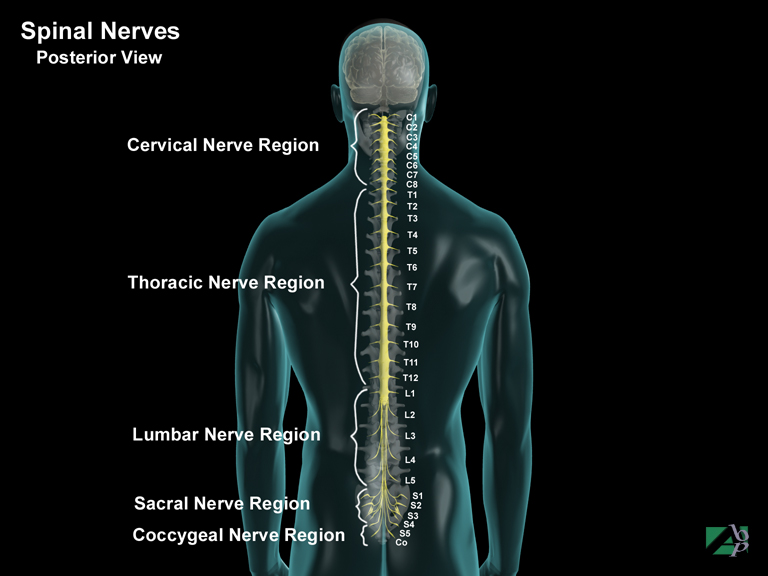
Thirty-one paired spinal nerves arise from the spinal cord. They are grouped according to the spinal region from which they emerge. There are eight cervical pairs, twelve thoracic pairs, five lumbar, five sacral and one coccygeal pair. Each spinal nerve consists of motor and sensory fibers. A dorsal and ventral spinal nerve root forms each nerve, which emerge from the spinal cord through openings (foramen) in the vertebral column at each of the intervertebral disc spaces. The ventral nerve root fibers are motor fibers while the dorsal root fibers deal with sensation. When the nerve roots merge they form mixed spinal nerves with both motor and sensory innervations. After the nerves pass through the intervertebral foramina they divide into four main branches called Rami. The rami then go to form plexuses by joining with rami from other spinal nerves. Plexus means "braid" which is what the network of nerves resemble. From the plexuses arise the peripheral nerves, which innervate the extremities. These peripheral nerves are named after the body region they innervate or the course they take. The principle plexuses formed by the spinal nerves are the cervical plexus, brachial plexus, lumbar plexus and sacral plexus
Spinal Radiofrequency Lesioning
Radiofrequency lesioning is technique, which uses a probe device that heats nerve tissue using radiofrequency signals that creates a nerve lesion thus interrupting the nerves impulses. It is a procedure used for the treatment of intractable pain
Spinal Stenosis
Narrowing of the spinal canal. It may be acquired, due to degenerative changes or congenital. It may be due to fractures, particularly burst fractures, and lesions such as tumors. Spinal stenosis is more common in the lumbar spine than other spine regions. Stenosis is often mistaken for disc lesions. The narrowing results in pressure being placed on spinal nerve roots
Spleen
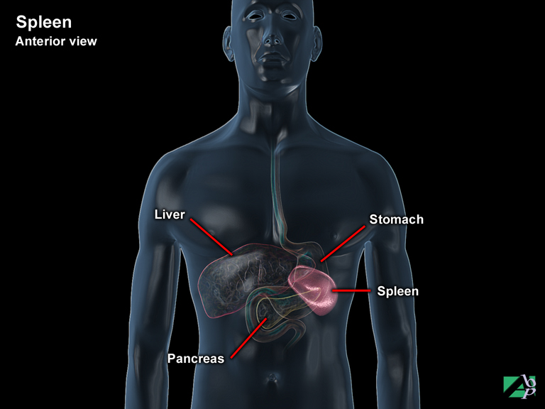
The spleen lies under the diaphragm on the left side of the abdominal cavity, behind the stomach and beneath the 9th, 10th and 11th ribs. The spleen acts as a filter against foreign organisms that infiltrate the bloodstream and also filters out old red blood cells from the bloodstream and decomposes them. During this process it also is involved in the metabolism of iron. As it filters defective red blood cells from the system, it salvages the iron for re-use in the creation of new cells. The spleen also acts as a blood reservoir; during stress or at other times when additional blood is needed the spleen contracts, forcing stored blood into circulation. The spleen also plays a role in the immune system producing substances that enhance destruction of foreign elements. Despite these functions, the spleen is not vital to life and individuals can function without it, although it is now thought that in its absence the individual is at a greater risk of infection and disease
Splenectomy
A surgical operation for the removal of part or all of the spleen
Splenius Capitis
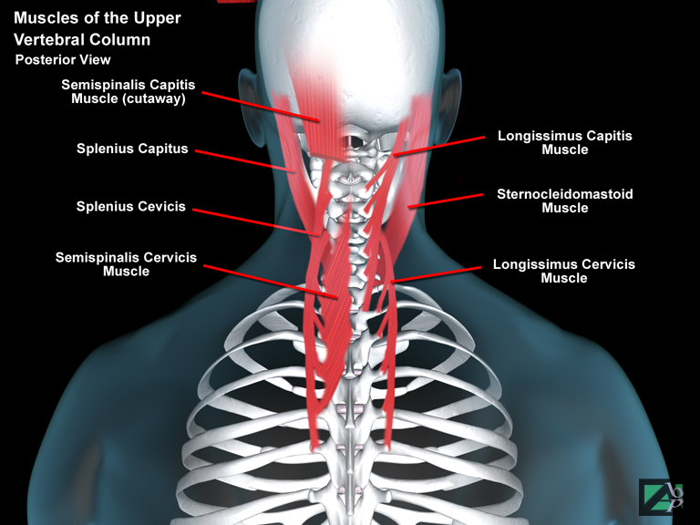
A muscle in the back of the neck originating from the spinous processes of T1-T3 and inserting onto the occipital bone. It assists extension and rotation of the cervical spine
Splenius Cervicis
A muscle in the back of the neck originating from T3-T6 and inserting onto the transverse processes of C1-C3. It assists extension and rotation of the cervical spine
Splenorrhaphy
A medical procedure, suture repair of the spleen
Spondylolisthesis
Spondylolisthesis is the forward displacement of one vertebral body on its immediate lower neighbor. The slippage is often classified according to the degree of displacement and four classes are used. A 1st degree slip is a displacement of one quarter of the anteroposterior vertebral body diameter while a 4th degree slip is a full diameter displacement. Any vertebra may be involved, but most commonly it is the fourth or the fifth lumbar vertebra. There are various types of spondylolisthesis and although traumatic spondylolisthesis is one type, trauma is rarely the cause of the condition and when it is the cause, an associated fracture of the neural arch will also be present. Trauma can however aggravate a pre-existing spondylolisthesis and cause further slippage of the affected vertebrae. Mostly the cause of spondylolisthesis is due to congenital reasons eg. failure of the neural arch to develop normally. Other reasons include bone infection, bone tumors and osteoarthritis.
Spondylolysis
Spondylolysis is a defect of the pars interarticularis of a vertebra. The pars is a narrow piece of bone that lies between the superior and inferior articulating processes of the vertebra. The defect may be found at any level but most commonly occurs in the lumbar spine, particularly at L5. Spondylolysis is also the most common cause of spondylolisthesis. The cause for the defect may be genetic, inherited or due to repetitive trauma and there is a strong association between spondylolysis and certain sports. Sports that have been associated include gymnastics, wrestling, diving, ballet dancing, pole vaulting, football, water skiing and weight lifting, baseball and cricket
Spondylopathy
Spondylopathy is a broad term used to describe any disease of the vertebra
Spondylosis
Spondylosis is the most common disorder of the cervical spine and results from chronic intervertebral disc degeneration. As degenerative changes develop and the disc loses height, the disc space narrows so that the annulus bulges beyond its normal confines. Osteophytes (bony outgrowths) develop circumferentially around the vertebral margins. Posterior osteophytes may protrude into the neural canal to produce neurological symptoms. Spondylosis is a gradual process and does not necessarily become symptomatic. Even if symptomatic, symptoms may be minimal. Pain is the primary symptom. In the cervical region the pain may be of the radiating type extending down one or both arms, or to the occipital and frontal regions and in the back in the scapula region. If nerve root or cord compression is involved paresthesia and weakness may also be symptoms. In the lumbar region spondylosis may produce sciatic like pain with pain radiating down into one or both legs. In the thoracic region pain may radiate around to the chest wall
Sprain
A sprain describes a stretching, partial tear or complete tear of a ligament that binds a joint together. The term is often used synonymous with strain but strictly speaking a strain is a stretching of a muscle, tendon or ligament. Sprains are sometimes classified by grade or described as being minor, moderate or severe. If graded a grade 1 sprain is equal to a minor sprain, one in which there is stretching of a muscle, tendon or ligament only, the structural integrity remains intact. Grade 11 sprains are the equivalent of a moderate sprain, in this type of sprain there is a partial tear. Grade 111 sprains are severe sprains. In this type of sprain there is a complete tearing of the structure
Squamosal Suture
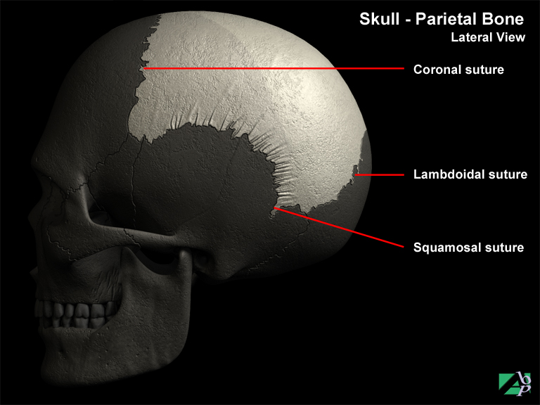
The skull is divided by tissue and cartilage called sutures, the squamosal suture divides the parietal and temporal bones
Stapedectomy
Stapedectomy is a surgical procedure to restore loss of hearing caused by the loss of normal vibration of the stapes, one of three bones in the middle ear called ossicles that transmit vibrations to the inner ear. The procedure involves removing the stapes and replacing it with a prosthesis, which conducts the vibrations in place of the stapes
Stapes
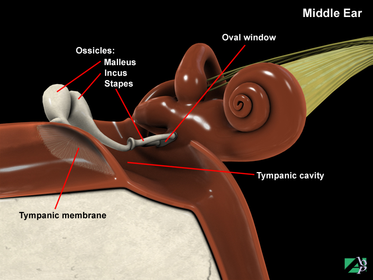
A bone of the middle ear referred to as an ossicle, the stapes and two other small bones, the incus and malleus, amplify and transmit vibrations from the eardrum to the inner ear
Stellate Fracture
A fracture where the fracture line(s) are starlike in appearance, they most commonly occur in the skull
Stent
A stent is a small flexible tube made of plastic or wire mesh implanted into blood vessels or ducts or hollow organs. It is most often used in vascular procedures, particularly with angioplasty. The stent serves to keep the structure open
Stenosis
Narrowing of the spinal canal
Sternal
Refers to the middle of the chest, the sternum area
Sternoclavicular Joint
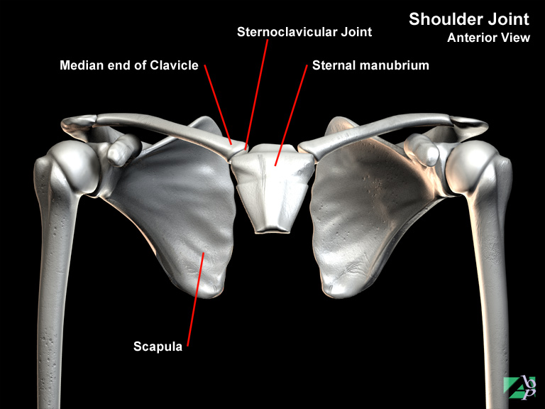
The sternoclavicular joint is formed where the clavicle (collar bone) connects with the manubrium of the sternum (the breast bone). Four ligaments provide the joint's movements: the anterior and posterior sternoclavicular ligaments, the interclavicular ligament and the costoclavicular ligament
Sternocleidomastoid
A muscle in the neck that originates from the sternum and clavicle and inserts onto the mastoid process (behind the ear) and occipital bone. It assists flexion and lateral rotation of the cervical spine and rotation of the head. It is innervated by the spinal accessory nerve
Sternum
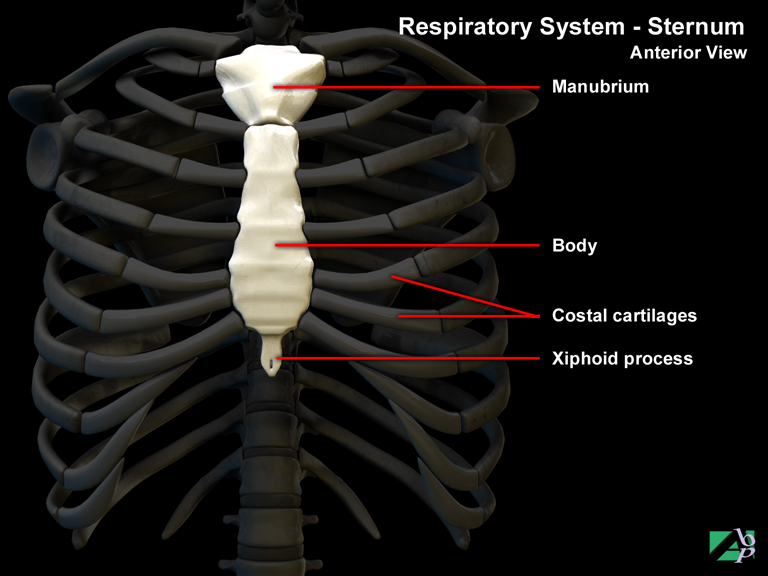
The sternum, or breastbone is made up of three separate bones. They are the manubrium, (the uppermost part of the sternum) the central body and the xiphoid process (the bottom of the sternum). The ribs attach to the sternum via costal cartilages to the costal notches of the sternum, which lie on the outer side of the sternum. At the top of the manubrium there are two more articulating notches called the sternal notch and the clavicular notch. The clavicular notches, which lay either side of the sternal notch, are for articulation with the clavicles. The manubrium is the attachment point for the first and second pairs of ribs. The sternum body is the attachment point for ribs 3 through 10. The xiphoid has no attachment with the ribs but is an attachment point for abdominal muscles
Stoma
An opening between two organs, it may be a natural opening or artificially created. An artificial stoma for example is a tracheostomy incision
Stomach
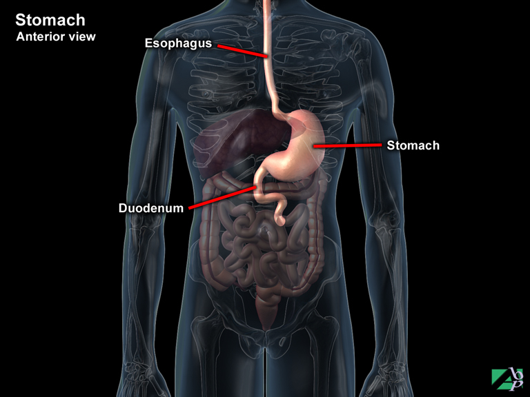
The stomach is a muscular sac like organ located between the esophagus and the small intestine; it serves as temporary receptacle for food. It has two openings. At the top where it joins the esophagus the opening is called the cardiac opening. At the bottom where it empties into the duodenum the opening is called the pyloric opening, or pyloric valve. The walls of the stomach are composed of four layers or tunics. The outermost layer is the peritoneal layer, the next underlying layer is the muscular coat, the next the submucous layer and the innermost layer is the mucous layer. The muscular layer is also composed of layers, three in all. The mucous layer or gastric mucosa is lined with many thousands of gastric glands that secrete a juice known as gastric juice, which is composed primarily of hydrochloric acid. The gastric juices convert the food into a semi-liquid state known as chyme, which is then passed into the duodenum. The stomach is capable of gross alterations in size depending on the position of the body and the amount of food inside. Food enters the stomach from the esophagus through the cardiac opening or sphincter and remains there until modified by the gastric juices. The duodenum opening, the pyloric valve, remains closed until this process is complete. The stomach functions under the autonomous nervous system and there is no control of it by the central nervous system
Strabismus
Cross-eyed
Strain
A stretching of a ligament
Stump Percussion
A method employed to treat phantom limb pain following an amputation. It involves striking the stump end
Subarachnoid Hematoma
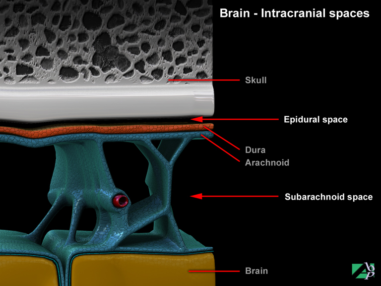
Traumatic subarachnoid hematoma occurs when blood leaks into the subarachnoid space from a ruptured artery or vein, this most often occurs concomitant with fractures of the skull. The subarachnoid space normally contains cerebrospinal fluid so when hemorrhaging does occur clotting does not take place and space occupying lesions don't occur
Subcapital Fracture
A fracture occurring just below the head of a bone
Subclavian Artery
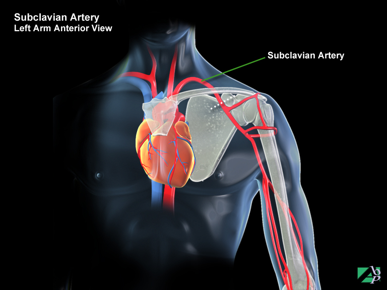
The subclavian artery lies deep below the clavicle. It is the major artery supplying blood to the upper extremity, although it is not truly an artery of the upper extremity. It has a number of branches, a vertebral branch (artery) that carries blood to the brain, a thyrocervical trunk that carries blood to the thyroid gland, trachea and larynx and an internal thoracic branch that descends into the thorax. As it passes into the armpit and enters the upper arm at a level with the 1st rib it becomes the axillary artery
Subclavius
A muscle of the shoulder that originates from the costochondral junction of the 1st rib and inserts onto the middle third of the clavicle. It depresses the clavicle
Subconjunctival
Subconjunctival refers to below the conjunctiva, the membrane that lines the outer surface of the eye and the underside of the eyeball
Subcostalis
A chest muscle that acts to depress the lower ribs
Subcutaneous
Refers to being below the skin
Subdural Hematoma
Subdural hematoma's occur between the dural membrane and the underlying arachnoid membrane. There are few attachments between the dura and arachnoid and, when hemorrhage occurs within this area a lesion may spread and occupy a large surface area. Subdural hematomas may be further classified as being acute, subacute or chronic. This classification is based on the time symptoms develop. Acute subdural hematoma produces neurological signs and deterioration in consciousness within hours of injury. Subacute subdural hematoma may not produce signs for days whereas chronic subdural hematoma may not demonstrate signs and be clinically diagnosed until weeks or months after injury
Subdural Space
The space or potential space between the dura mater and the arachnoid membrane. The dura mater is the outer layer of the membrane that covers the brain and spinal cord. The arachnoid is the next layer
Subluxation
A partial dislocation
Submucous
Lying below a mucous membrane. Mucous membrane exists in the mouth, throat, abdominal cavity etc
Subscapularis
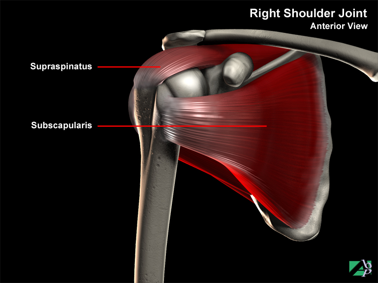
A shoulder muscle originating from the scapula and inserting onto the lesser tubercle of the humerus. It assists medial rotation of the arm and provides shoulder stabilization. The subscapularis nerve innervates it
Subtalar Joint
The joint formed by the talus and the calcaneus in the foot
Superficial
Pertaining to or relating to the surface of the body, in reference to an injury it denotes a minor or trivial injury
Superior
Refers to being above, lying above
Superior Gluteal Nerve
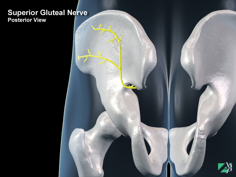
This nerve is found in the buttock. It provides motor activation to the abductor muscles of the thigh
Superior Mesenteric Artery
The superior mesenteric artery, which is a branch of the abdominal aorta supplies blood to the ascending colon, duodenum, jejunum, ileum and appendix
Superior Oblique
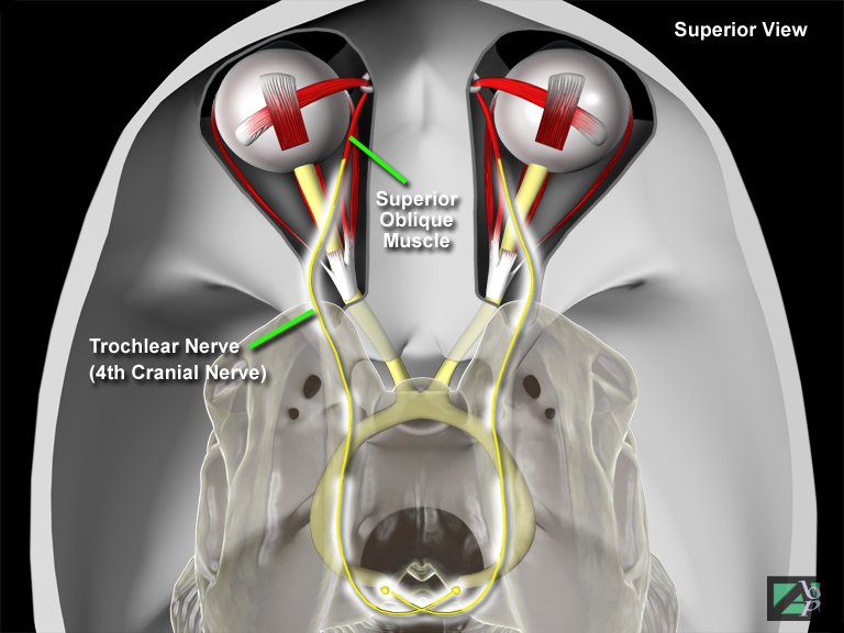
An eye muscle originating from the sphenoid bone and attaching to the back of the sclera of the eyeball. It depresses the eyeball. It is innervated by the trochlear nerve, one of the cranial nerves
Superior Rectus
An eye muscle originating from the orbital area and inserting onto the top of the sclera of the eyeball. It elevates the eye. It is innervated by the oculomotor nerve, one of the cranial nerves
Superior Vena Cava
The superior vena cava is a large vein that carries blood from the upper part of the torso to the right atrium of the heart
Supinator
A muscle of the forearm originating from the elbow and proximal ulna and inserting onto the shaft of the radius. It assists supination of the forearm
Supracondylar
Refers to above a condyle. A condyle is a rounded eminence on a bone; supracondylar refers to being above the condyle, as in supracondylar fracture, a fracture occurring above the condyle
Supraorbital
Being above the orbits, the eye-sockets
Supraorbital Foramina
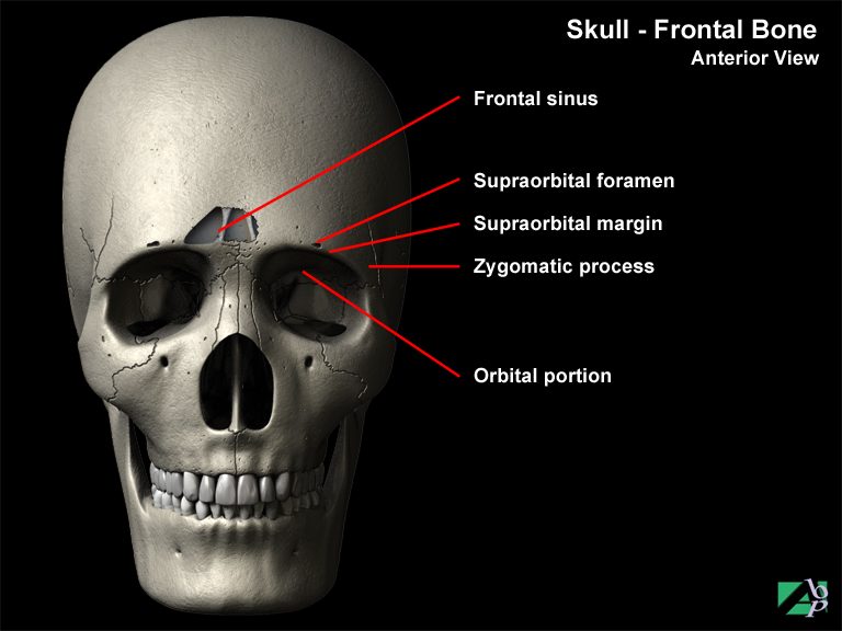
An opening along the supraorbital margin, part of the frontal bone (the eyebrow area) through which nerves and blood vessels pass
Supraorbital Margin
Part of the frontal bone, it is the bony ridge over the orbit, the eyebrow area
Supraspinatus Muscle
A muscle of the shoulder originating from the scapula and inserting onto the greater tuberosity of the humerus. It assists abduction of the arm and helps stabilize the shoulder joint
Sural Nerve
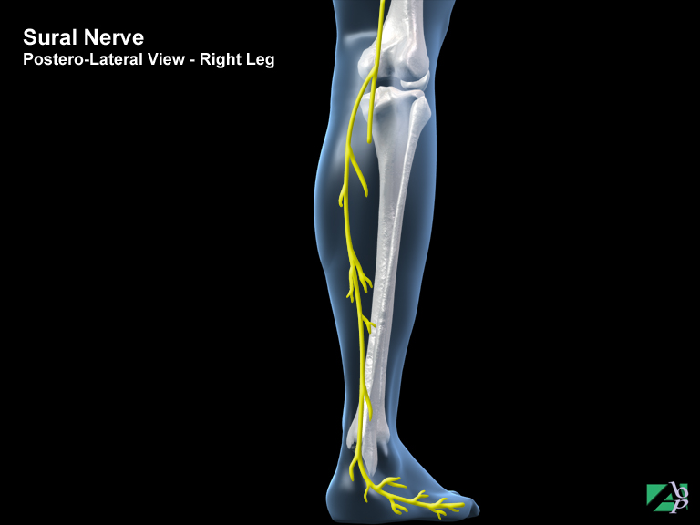
This is a sensory nerve that provides sensation to the inside and back of the leg and the ankle
Surgical Emphysema
Surgical, subcutaneous and traumatic emphysema is the presence of air or gas within the tissue beneath the skin. It can be localized or affect the whole body. It is usually found on the upper torso or neck and can be caused among other things by air leaking from the lungs following trauma or surgery
Symptomatic
Producing symptoms, a term used to describe an injury, disease or other medical condition that is causing or producing symptoms, as opposed to asymptomatic, which describes a condition present but one not producing symptoms
Syndesmosis
A type of joint where ligaments hold the bones together alone
Syndesmotic Ligament
A ligament in the ankle that helps support the ankle joint
Synovectomy
Synovectomy is the partial or complete surgical removal of a synovial membrane of a joint. It is often performed on individuals with severe arthritis. The wrist, knee and fingers are the joints most frequently involved. The synovial membrane lines joint cavities and secretes synovial fluid to lubricate and provide nourishment for the joint.
Synovectomy may be performed by open surgery or arthroscopically. If it is performed arthroscopically, enter both the synovectomy and the arthroscopy
Synovial Membrane
A membrane surrounding (lining) joint cavities, the membrane produces synovial fluid, which acts as a lubricating agent for the bones of the joint
Synovitis
Inflammation of a synovial membrane